9: Cervicofacial Rhytidectomy

9 Cervicofacial Rhytidectomy

A textbook on cosmetic facial surgery has the power to influence surgeons, surgeries, and outcomes in positive or negative ways. In giving advice, teaching, or showing how a given procedure is performed, its author is in a position to share a lifetime of learning, teaching, and trial-and-error experience with the book’s readers. Some editorialization relates to hows and how nots distilled from days past, new technology, and opinions that are solely those of the author. All readers must keep in mind that there are many successful means of achieving aesthetic outcomes for all procedures, and there is rarely one way to do any procedure.

My goal in writing this chapter is to share my experience of facelift surgery and focus on what I believe are sound, safe, and effective means of achieving predictable outcomes—tenets based on performing almost 700 facelifts. I have written numerous articles on facelift surgery, so the chapter will occasionally quote my own material, but it offers in-depth discussion of facelift surgery from the consult to the thank-you note.

Facelift surgery, or cervicofacial rhytidectomy, remains the ultimate rejuvenative cosmetic procedure. No other procedure can produce results patients can clearly see and appreciate 24 hours a day and do so much to boost confidence and self-esteem. Contemporary facelift surgery is a combination of art and science and requires significant experience to become proficient. Many subtle, technique-sensitive operative steps and nuances must be appreciated by the surgeon to provide predictable results with low complications. I feel this is one of the most challenging procedures to learn.

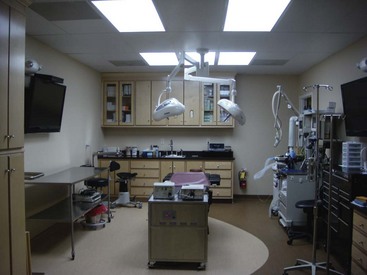
Like so many other things in life, the basics of facelift surgery are relatively straightforward, but the key to success is in the details, and the details take years to appreciate. Any given detail of the procedure may work well for some surgeons but not so well for others. Facelifts can be taxing; they can take from image to 4 hours. Most frequently, I am doing concomitant procedures such as blepharoplasty, brow lift, laser resurfacing, chin and cheek implants, and so forth along with the facelift. This sometimes translates into 5- to 6-hour cases. This has required me to make many changes in my office. I had our facility accredited by the Accreditation Association for Ambulatory Health Care (AAAHC) and equipped hospital-caliber operating rooms (Figure 9-1).

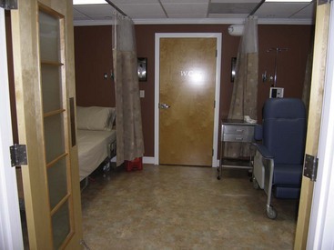
My original discipline is maxillofacial surgery, so I am well trained in ambulatory outpatient anesthesia and licensed in my state to perform intravenous (IV) sedation and general anesthesia. For many short procedures on healthy patients, we are able to use IV sedation with tumescent anesthesia. For longer cases or those on older or medically compromised patients, we use nurse anesthetists or physician anesthesiologists. I have also built a hospital-grade recovery suite suitable for ambulatory outpatient anesthesia but capable of overnight stay with nursing care (Figure 9-2). I have privileges at several local hospitals and surgery centers but rarely use them (with the exception of cases of medically compromised patients), opting for the convenience of my office surgery center. Although I believe I currently have a state-of-the-art facility, this did not happen overnight. Over the past 12 years, I have upgraded from relatively humble but safe facilities as the practice grew and the patient base expanded.

Facelift History

The scientific literature is replete with descriptions of early facelift techniques from the early 20th century that primarily detailed skin tightening and excision.111 In 1973, Skoog presented a technique of elevating the platysma muscle without detaching the skin.12 In 1976, Mitz and Peyronie described the superficial musculoaponeurotic system (SMAS),13 and later other authors described techniques of SMAS plication and imbrication.1416 By the late 1970s and mid-1980s, a combination of complete platysmal muscle transection, plication of medial borders, and pulling laterally was presented as the way to get the “best result.”17,18 Patient complaints, complications, and overoperated necks occurred, and many of these techniques were abandoned.19 Deep-plane and subperiosteal techniques have been described by multiple authors,2022 as well as endoscopic approaches.23

Many other techniques have been described, and some surgeons have gone as far as “inventing” new techniques and patenting catchy names to describe some minimally invasive facelift that is supposedly revolutionary. For the most part, these “new” techniques are a rehashing of previously described techniques (Figure 9-3). When reviewing the literature, it should be kept in mind that just because a technique has been published or promoted does not mean it is better or safer. It will be mentioned many times throughout this chapter that patient safety and outcome are testaments to “the best” facelift technique. Some techniques may work well in the hands of a given surgeon, and if they provide safe procedures with great outcomes, then they cannot be criticized. For the novice surgeon, however, it is preferable to begin with safe and predictable techniques before advancing to more complex procedures. Walk before you run!

At some time, the surgeon and patient are going to have to agree on what type of facelift is preferable for the given situation, and herein can lie a contemporary problem: many surgeons who are just learning facelift surgery may fixate on one of the “miracle lift” procedures promoted as being “easy, done under local anesthesia, no bandages, 48-hour recovery,” and the like. Since this type of lift is easier to learn, the novice may attempt to apply this single type of lift to all types of patients. These small lifts may work well on younger patients, but on older patients or those with more advanced aging, conservative techniques will not produce an adequate or lasting result. Patients with bigger problems need bigger procedures.

There is a time, place, and patient for short-cut procedures, but not all facelift patients can be painted with that brush. Someone once said, “Before learning the tricks of the trade, learn the trade,” and this sums up my point. There is no one-size-fits-all operation for facelifting. Each patient and operation must be customized specifically for the situation at hand. On a regular basis, I see unhappy patients who drove hours to get some trendy “franchise” lift that sounded too good be true, and they now present to my office with results that are less than promised and promoted. Almost unanimously, they spent more money getting half a facelift at a franchise than they would have spent getting a “real” facelift in my office.

Facelift Anatomy

Facelift surgery and anatomy are inseparable, good surgeons are great anatomists, and facial anatomic expertise is imperative to performing competent facelift surgery and avoiding complications. With the unique anatomy of the face, it is impossible to cover everything in this chapter; we will discuss the most relevant anatomy as it applies to the procedure.

The first layer concerned in facelift anatomy is obviously the skin. The dermal plexus of blood vessels nourishes the skin (and facelift flap) and must at all times be respected. The handling of lipocutaneous flaps in cosmetic facial surgery should be as gentle as other procedures in which the vascularity of the flap is essential to its survival. When dissecting a facelift flap, several millimeters of fat is left adherent to the dermal undersurface to protect this area and enhance flap viability. A good general rule is that there should be fat on both sides of the facelift flap.

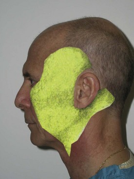
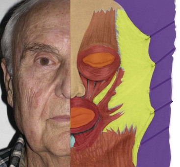
The next layer is the subcutaneous layer (Figure 9-4). This is the layer of fat that is intimate to the dermis superficially and also with the deeper SMAS. The subcutaneous layer is basically a safe layer in the face and can be undermined in the subcutaneous plane without damage to significant anatomic structures. This layer is of varying thickness depending upon the location and patient. This layer also becomes thickened over the malar region, where dissection can be tenuous. This tissue is attached to the malar periosteum by retaining ligaments that run from the underlying periosteum through the malar pad and insert into the dermis. This area is very tenacious and because of its fibrous nature provides resistance when dissecting; it is referred to as McGregor’s patch. Brisk bleeding is also frequently seen in the area due to accompanying vasculature.

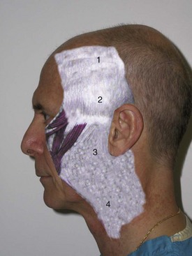
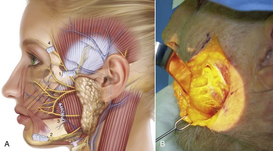
The third layer is the SMAS layer. This layer separates the overlying subcutaneous fat from the underlying parotid-masseteric fascia and facial nerve branches (Figure 9-5). The SMAS layer, as described by many leading authors, is said to be continuous with the galea in the scalp, the temporoparietal fascia in the temples, and the superior cervical fascia in the neck. This structure was initially described by Mitz and Peyronie as muscular and fibrous tissues and termed the superficial musculoaponeurotic system.13 They stated the SMAS exists in the parotid and cheek region and invests the muscles of facial expression. In their original article, the authors state, “To us, the SMAS appears to be a fibromuscular network located between the facial muscles and the dermis, one which covers the facial motor nerves.” They note that the SMAS separates two layers of fat in the face into superficial and deep layers. Their work also implied that the SMAS was continuous with the platysma.

Various authors contest Mitz and Peyronie’s finite description of the SMAS and related layers, and some surgeons do not agree that there is any such thing at all as the SMAS. Jost and Levet24 refuted the work of Mitz and Peyronie and dispute their findings. In this study, the authors combine embryology and comparative anatomy of other mammals as well as cadaver dissections and summarize that the SMAS as described previously is incorrect. These authors describe the SMAS as a double-layered system. The first layer is the superficial system which is consistent with primitive platysma and includes parotid fascia and has no direct bone insertion. In addition, they state that a deep system exists which includes the sphincter coli profundus. This group of muscles in the perioral region does possess bony attachments. These authors also contradict Mitz and Peyronie and state that the SMAS is not continuous with the platysma, but rather it is the parotid fascia that is continuous with the platysma. Additionally, they comment that the parotid fascia is the uppermost part of the primitive platysma, which is a muscle that has undergone fibrous degeneration. Applying this to facelift surgery, these authors suggest dissection deep to the parotid fascia.

In an additional article denying that the SMAS exists as originally described,25 Gardetto et al. state that according to the work of Mitz and Peyronie, “no SMAS exists in any of the described regions that agrees with the basic criteria of Mitz and Peyronie.” This paper corroborates through comparative anatomy and fresh cadaver dissection studies that the “SMAS does exist in the parotid region, where it is thick but becomes very thin and discontinuous in the cheek where it is impossible to identify or dissect it, even with microsurgical technique.” They further state that “there is no evidence of continuity of the SMAS and the temporal fascia but in contrast, in the lower eyelid there is continuity with the lateral portion or the orbicularis oculi muscle.”

Having said all this, one can call the fibromuscular layer anything they wish (or deny its existence). Most surgeons will agree it separates the subcutaneous fat from the underlying parotidomasseteric fascia. When a surgeon states that they pick up the SMAS, in reality they are picking up adipose tissue of the subcutaneous fat and fibromuscular tissue above the parotidomasseteric fascia. This is what is frequently plicated, excised, imbricated, and otherwise managed in facelift surgery (Figure 9-6). Whether you agree about the embryology and histology, we will refer to this layer as SMAS throughout this text. All motor nerves providing movement of the facial muscles are deep to this plane. When this layer is stretched or pulled, it pulls on the mimetic muscles and basically moves the entire lateral face in the desired vector. This makes sense; it is said that the physiologic function of the SMAS is to more effectively transfer mimetic muscle movement to the facial skin by septae that extend from the SMAS to the dermis. In theory, this would allow the face to move more as a unit (as opposed to each individual muscle), thus making expression more efficient.

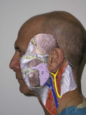
The fourth surgical plane commonly involved in facelift surgery is the sub-SMAS plane (Figure 9-7). This surgical plane contains the facial nerve motor branches and the parotid duct and is best avoided by novice surgeons. The facial nerve branches are deeper in the parotid gland until they emerge at the anterior border of the gland and cross the masseter muscle. The parotidomasseteric fascia is the facial layer that overlies the parotid gland and masseter muscle; when operating superior to this layer, the facial nerve branches are generally protected. Just as the SMAS is an extension of the superficial cervical fascia, the parotidomasseteric fascia is an extension of the superficial layer of the deep cervical fascia into the face and the deep temporal fascia above the zygomatic arch. The novice surgeon should not violate the parotidomasseteric fascia, although many seasoned surgeons perform deep plane dissections beneath the layer routinely. Obviously the deeper and more anteriorly the dissection proceeds beneath this layer, the greater the chance of injury to the facial nerve branches. There is a sub-SMAS loose areolar tissue plane that extends from the anterior border of the parotid to the anterior border of the masseter. Blunt dissection in this plane allows the deep plane facelift dissection to proceed safely even though it is almost intimate with the underlying facial nerve branches.

As the facial nerve branches course anteriorly, they overlie the buccal fat pad then innervate the mimetic muscles. Besides the facial nerve branches and parotid duct, the buccal fat pad and facial artery and vein also lie in the plane under the parotidomasseteric fascia.

The relationship between the parotid gland and the facial nerve branches will be referenced many times in this text, and the reader should keep in mind that dissection over the parotid gland is safe, but the facial nerve branches are at risk as they exit the gland and traverse the masseter. Staying over the parotid and above the parotid fascia is the safest anatomic area for novice facelift surgeons. So long as dissection stays superficial to the SMAS and platysma, the motor nerves are protected.

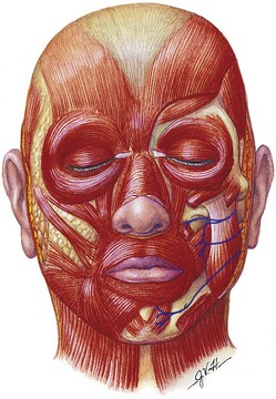
One reason the facial nerves are protected in deep plane dissection as they branch in the anterior face is that most nerves innervate the muscle on its posterior surface. The only muscles of facial expression that are exceptions are the levator anguli oris, the buccinator, and the mentalis, which are all innervated on their anterior surface (Figure 9-8).

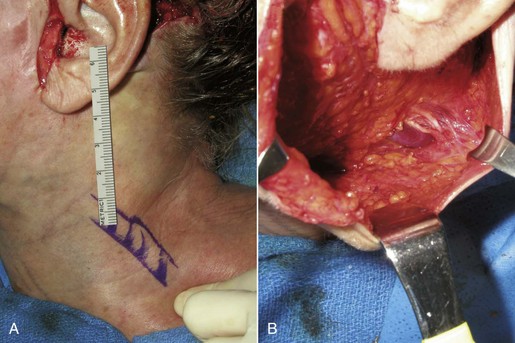
Several other structures are frequently encountered in facelift surgery and are vulnerable to injury. The sternocleidomastoid muscle is significant because the external jugular vein and greater auricular nerve are intimate and traverse this muscle in the midlateral neck (Figure 9-9). The greater auricular provides sensory innervation to the earlobe and lateral cheek. The external jugular vein and greater auricular nerve lie deep to the SMAS and are protected as long as the dissection remains in the subcutaneous plane. The subcutaneous tissue becomes very thin in this part of the neck, and the dermis is sometimes intimate to the fascia of the sternocleidomastoid muscle, which can put the nerve and vein in jeopardy.

The youthful face is in part suspended by retaining ligaments that support the skin over the facial bones. These ligaments become lax with age and in part are responsible for the sagging of facial tissues. This connective tissue can run from bone to dermis (osteocutaneous ligaments) or from soft tissue to dermis (cutaneous ligaments). The exact name and location of these ligaments are described differently by various authors but collectively can be appreciated during facelift dissection, where they can be felt (fibrous tissue difficult to bluntly dissect) or visualized directly. Figure 9-10 shows a rendition of the approximate position of these retaining ligaments.

Patient Selection

In theory, most patients in their fourth decade are candidates for some type of facelift. Younger patients may seek consultation for minor jowling and neck changes and be candidates for minimally invasive rhytidectomy, whereas older patients may require larger procedures with other concomitant mid- and upper facial cosmetic surgery correction.

One problem is that the word facelift conjures different ideas for each patient. Some patients think a “full” facelift includes eyelid and brow surgery or other procedures. Likewise, I have had postoperative facelift patients complain that they still have wrinkles around their mouth. The first goal of facelift consultation is to explain the procedure and educate the patient about what the procedure does and does not do. Should the case come to fruition, I have a special consent, “What Your Facelift Won’t Do.” This is important information; patients may assume that all their wrinkles will be improved or that their result will last forever or that they may look 20 years younger.

Many patients are surprised when I explain that a facelift is a procedure primarily for the lower face. It is not uncommon for patients to present and request “just a little tuck to pull back my cheeks.” They place their fingers on their cheeks and reset their cheeks, jowls, and neck (Figure 9-11, A). It must be explained to these patients that no procedure exists that corrects aging in that vector. By pulling the skin in a more lateral direction, a more representative correction can be explained (see Figure 9-11, B). A more accurate means of illustrating the potential changes from cervicofacial rhytidectomy is to place the patient supine and have them look in a mirror while elevating their chin. By removing gravity from the equation, the brows and midface are elevated, and the jowls and neck are vastly improved. I find this to be a relatively accurate prediction of the final lower face result (Figure 9-12). Obviously, digital morphing can be performed, but oftentimes the patient will set their sights on the morphed postop image and express unhappiness if that picture is not duplicated by the surgery.
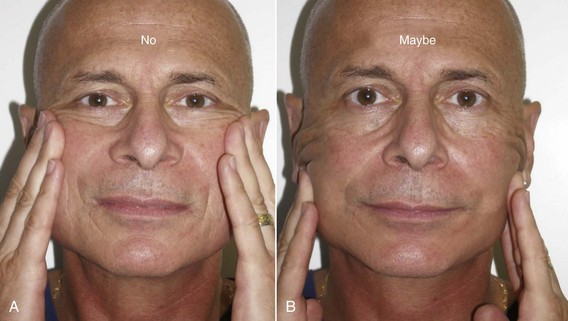
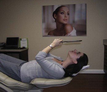
I also show the patient before-and-after pictures of actual facelifts I have done and refer them to my website for more pictures and explanations. I offer them the opportunity to speak with other patients who also have had similar procedures.

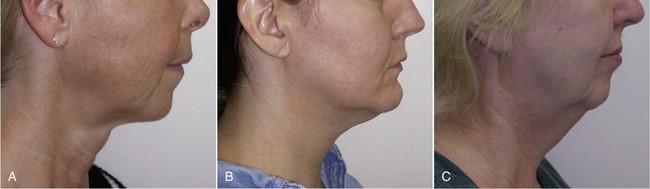
Many classifications exist for grading aging changes, but I truly believe it is easier to think in terms of small, medium, and large (Figure 9-13). As stated earlier, a facelift is primarily a procedure for the lower face. No other procedure can address the jowls and sagging neck. Many shortcuts exist, such as liposuction and submentoplasty, but in the average patient with noticeable aging changes, cervicofacial rhytidectomy is a time-proven procedure to provide natural and lasting results.

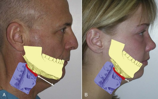
Patients with large neck and jowl changes are poor candidates for the novice surgeon. Beginners should initially concentrate on thinner patients with mild aging and normal chin position. When considering candidates for rhytidectomy, chin and submental position is also of significant consideration. In patients with retrognathia or microgenia, the final result will not be as dramatic unless chin augmentation with implants or osteotomy is performed. Even more important is the position of the hyoid bone and associated muscles of the submental region. In patients with a hyoid position and laryngeal anatomy that is superior and posterior, the skin can be pulled to form an acute angle that approximates 90 degrees (Figure 9-14, A). Patients with this type of anatomy are better candidates than those with inferior and posteriorly situated hyoid bones and submental anatomy (see Figure 9-14, B). The novice surgeon must realize these implications. On patients with anatomy consistent with Figure 9-14, B, chin augmentation can improve the final outcome, but these patients must realize that their particular anatomy limits the result when compared to a patient with normal hyoid and submental position. I keep a picture of Figure 9-14 in my evaluation rooms to explain this concept to prospective patients.

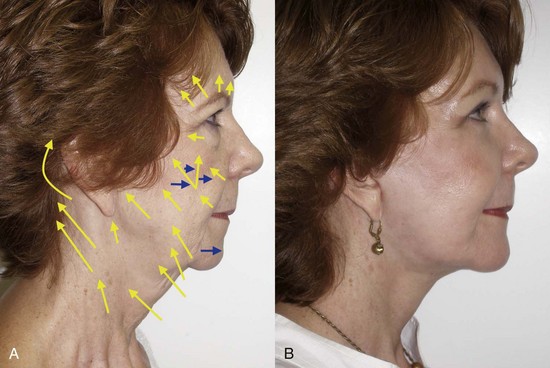
Correct diagnosis of the patient’s condition and how best to improve it can be a challenge for the novice surgeon. Appreciating the vectors of facial aging (described in Chapter 1) can shed light on an orderly manner for the diagnosis and rejuvenation of the aging face. Figure 9-15 shows a rendition of a patient with panfacial aging and the respective vectors that need correction. Having a mental picture of this can assist the surgeon in developing an appropriate treatment plan. Actually showing a picture similar to Figure 9-15 to the patient can improve their understanding of the proposed surgery.

Determining Status for Potential Cosmetic Facial Surgery

During facelift consultation, I give patients four criteria required to be a candidate in my office: health, psychological stability, recovery, budget.

Health

The patient must be in adequate health for the planned procedure. Although most patients are candidates for some type of rhytidectomy procedures, it is not uncommon that they have systemic problems such as hypertension, cardiovascular disease, diabetes, hyperlipidemia, arthritis, osteoporosis, and other conditions. Patients are living longer and taking more medications, so the medical status may be complex. Most of these patients are candidates for cosmetic surgery if their diseases are controlled and/or medically managed. Patients taking anticoagulants or medications that affect platelet function present special problems and may require intense medical management. The more problems a patient has, the more risky surgery and anesthesia can be. There comes a point at which the risk of surgery outweighs the benefits of looking younger. In and of itself, age is not a contraindication to facelift surgery. I have treated 78-year-old patients who were healthier than some of my 50-year-old patients. Older patients, even healthy ones, may have decreased function or not tolerate extended surgeries and recoveries as well as younger patients, and the treatment plan must be tempered. All my facelift patients are required to have a history and physical and electrocardiogram from their physician, as well as age-appropriate laboratory studies. All patients get coagulation studies and HIV and hepatitis C screening. Chest x-ray, pulmonary function tests, and cardiac workup, including stress test and echocardiograms, are performed if suspicion exists concerning specific health status. Tolerating the surgery is obviously a concern, but tolerating the anesthesia can also be critical. When patients present with questionable cardiac status, I refer them to a cardiologist and inquire about the following:

For all patients, it is imperative to have a written document from the needed health providers that the patient is stable and cleared to undergo anesthesia and surgery. In the event of anesthesia or surgery problems, this omission is one of the first things a plaintiff’s attorney would seek.

Smoking, alcohol, or drug abuse are lifestyle factors that could influence surgery and anesthesia as well. Smoking is a particularly common factor encountered. Some surgeons refuse to operate on smokers. I practice in Richmond, Virginia, where smoking is common. I would estimate that 25% of my patients smoke cigarettes, and this is not a contraindication to facelift surgery. I am somewhat more cautious with smokers and avoid very large dissections. I will usually perform concomitant laser resurfacing but treat the flaps with much less fluence. For extremely heavy smokers (two packs per day), I don’t place as much tension on the flap or perform simultaneous laser. The average smoker will report their habit as a pack a day, and these patients generally fare well. Patients who smoke two packs per day, in my experience, have increased airway and anesthesia-related problems. All smokers must sign a smoker’s consent for all cosmetic surgery procedures. This consent details the facts of compromised healing and other negative factors that are particular to smokers. Another problem is patients underreporting the extent of their smoking or denying smoking at all when they are actually smokers. A simple, low-cost saliva nicotine test, NicAlert, (Nymox Corp., Hasbrouck Heights, NJ), is available and could be used to truly determine if a patient smokes or otherwise abuses tobacco. Although many surgeons and anesthesiologists recommend smoking cessation before surgery, I usually do not. I have had several patients who used their facelift as reason kick their habit, but it is rare. I have also seen patients decompensate from the physical and emotional aspects of nicotine withdrawal and complicate their pre- and postsurgical experience. Patient death with any surgery is a horrible circumstance.

If it should occur with elective surgery, it is even more disastrous. Besides the loss of life and the sorrow of the family, surgeon, and staff, an event like this could end a practice. As more and more cosmetic surgery is performed in the ambulatory office outpatient setting where there is less emergency help and trained personnel, meticulous health screening is imperative. All the required preoperative details of lab tests, histories and physicals, and similar screening can be very time consuming and fall between the cracks. Strict staff monitoring of these details is invaluable and can be lifesaving.

Psychological Stability

Patients should be requesting cosmetic surgery for the correct reasons, but emotional stability is difficult to ascertain because it is difficult to quantify. Panfacial rejuvenation involves a significant recovery process, and patients who seem psychologically strong preoperatively can decompensate emotionally in the recovery period to the point at which they need psychiatric assistance.

A number of warning signs exist that should be taken into serious consideration when deciding whether a patient is a suitable candidate for any cosmetic surgery. Frequently the office staff is quite astute at picking up on the potentially unsuitable patient, and we have witnessed common scenarios that serve as red flags. It is not uncommon for this type of patient to be difficult to deal with throughout the entire process. They are pushy and demanding to the receptionist when scheduling, they exhibit the same behavior with the evaluation staff, they are resistant to the inconvenience of having a medical workup and labs, and they fight with the financial coordinator. The best way to handle such a patient is to not treat them. We have all had the experience of unpleasant dealings with patients we know we shouldn’t have accepted. It is not a comfortable position to be in. Learn to say no. Telling a patient you feel you can’t make them happy is an easy and polite means of discharging them preoperatively.

Over the past 25 years, I have seen repeating red flags for cosmetic surgery:

This list does not necessarily mean I won’t operate on such patients, but any of the above and especially a combination of the above should serve to alert the surgeon and staff of a potentially difficult experience.

Recovery

Patients must be able to take time away from work or play commensurate with the procedure(s) considered. Everyone is busy, and with more women in the work place, it is difficult to balance cosmetic surgery and work. One of the biggest mistakes a surgeon can make is to undersell a recovery. If you tell a patient they will recover in a week and it takes 2 weeks, they can be furious. If you tell them it takes 3 weeks and they recover in 2 weeks, you will be a hero. Many of the aspects of surgery, such as incisions, sutures, anesthesia, bandages, and so forth, can be objectively and accurately described. Recovery, on the other hand, is very subjective. I have, on several occasions, performed the same procedure on identical twins, and one twin did great while the other twin had unusual swelling and bruising. It is impossible to guarantee exact recovery, and the surgeon must rely on the mean recovery of a procedure or procedures. I tell facelift and laser patients that the average patient can return to work or social situations at 2 weeks. I explain to them that on the bell curve, some patients may look great at 9 days and others may still be bruised at 3 weeks. The surgeon should always err on the high side; if the patient is given a range of 6 to 14 days, 6 days is the only number they will remember. All of this needs to be covered in the consent and presurgical material issued to the patient. I give patients the option of a Friday surgery which gives them a weekend, a full work week, and another weekend to recover. This is sufficient for selected surgeries, but I tell them that no surgeon can guarantee a specific recovery. If they are having a facelift before a big event like a child’s wedding, a reunion, or an important vacation, I suggest 4 to 6 weeks for recovery. I also explain that the recovery process in actuality takes about 90 days, and they will see positive changes throughout this period.

Preoperative Consultation Workup

Facelift surgery, especially when combined with ancillary procedures, is very detailed. There are many specifics such as preoperative workup, prescriptions, and postoperative and follow-up appointments. This can be very logical for the office that does it every week but very complicated for the patient who does it once in their life. It helps to have very detailed instructions and a check-off list for your staff to follow the progress of the communication process.

As stated earlier, all facelift patients are required to have a physician history and physical, ECG, coagulation profile, complete blood count, and HIV and hepatitis C screening. We ask that this information be back to our office at least 2 weeks before the surgery, and inevitably we are pushing the deadline. The staff must stay on top of the progress; otherwise there is no time to work up abnormal studies before surgery. The preoperative appointment generally takes 45 to 60 minutes, most of which is attended by the staff. At the preoperative appointment, patients are given their prescriptions and a prescription for compression stockings. They are also given instructions to use an antibacterial bath soap for body and shampoo the night before and the day of surgery. They are given their postoperative appointment schedule and also take care of their financial arrangements. Our policy is that half the surgical fee is due 2 weeks before the procedure, and the other half is due on the day of surgery. All consents are signed at this appointment, and the doctor is present for any final questions. Preoperative digital photographs are taken at this appointment, traditionally with the patient wearing makeup. A second set of preoperative digital photographs are taken on the day of surgery without makeup. These images are essential for medicolegal records and marketing and can prove integral to resolving conflicts with unhappy patients.

Finally, the patient’s home support is discussed. They are informed that they need a responsible adult to monitor them the first several days postoperatively and drive them to my office on the first postoperative day. If there is no caregiver, they are given the option of private duty nursing at their home or at my office on the night of the surgery.

It is not uncommon for patients to be quite nervous at this appointment, since they have read multiple consents; some patients consider canceling surgery. I explain to them that significant complications are rare, but no surgeon can guarantee complication-free surgery. I also tell them that if they do have a complication, I will stand by them and do everything possible to correct it.

The topic of revision surgery should be addressed verbally and in the consent. What will happen if the result is not as planned? Will the surgeon perform revision surgery, and who will pay for it? Once in a while, the best-executed facelift will relapse earlier than expected. It can occur from the patient’s inherent problems with excess skin elasticity, weight gain or loss, or from surgical relapse. My personal policy is to perform free of charge, reasonable surgical revisions that relate to the initial result, such as small areas of skin or fat excess or scar revision. I do not apply a surgical fee but do require a nominal materials and anesthesia fee. From time to time, a patient will expect free revision surgery for a situation that is within normal limits for the procedure performed; those patients can be dealt with in a different manner. Again, I always try to remember that “the customer is always right.” If these topics are discussed preoperatively, they are generally easy to negotiate after surgery, but if the topic is not discussed until after surgery, communication can be uncomfortable, awkward, and emotional.

This should all be in the consent. “If it is not written down, it didn’t happen” is another rule to remember.

When the patient walks out the door at this appointment, all the i’s should be dotted and the t’s crossed. The patient may be apprehensive but reassured and medically, physically, and emotionally prepared for the upcoming procedures.

Facility

Although many surgeons perform facelift surgery in a hospital or accredited facility, it is not an absolute requirement. The supreme importance is patient safety. If the surgeon is performing surgery with local or tumescent anesthesia or light IV sedation, then the actual facility is less important. With IV sedation, one must have the appropriate emergency equipment: vital sign monitors, oxygen, instruments for emergency airway placement, medications for such anesthetic emergencies as anaphylactic shock, allergy, hypertension, hypotension, and the like.

Many novice facelift surgeons begin with office-based surgery performed in their ambulatory office surgical suite. Although most surgeons perform facelifts with full sterile technique, some doctors operate in a “clean” surgical environment. Postoperative infections are rare in head and neck surgery, and I have not experienced a significant one after thousands of cosmetic facial procedures, regardless of sterile versus clean technique. This is especially true for minimally invasive procedures. I personally perform facelift surgery in a full scrub, sterile environment as done in the hospital. Being gowned is important for the surgeon and staff to be protected from potentially bloody procedures. Most practitioners already have minor surgery suites in their offices that can easily accommodate basic rhytidectomy. If longer cosmetic cases such as facelift with other procedures become a significant portion of one’s practice, a more formal and elaborate setup is required.

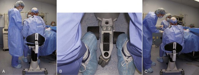
There is no substitute for a formal operating table, and these can be purchased refurbished. Alternatively, a dental or minor surgical chair can suffice. Having the ability to position patients laterally (airplane) can be of great assistance, especially with older patients with limited neck mobility which makes positioning the head difficult. This prevents the surgeon from bending over and working in a hole. An additional item that has become personally indispensable is an operator stool that can be vertically repositioned (Figure 9-16). Cosmetic facial surgery and especially facelift surgery requires many different patient and surgeon positions, and having the ability to pump up or lower the stool makes it much easier to perform seated surgery. I begin in an elevated position but frequently “drop down” to look under the preauricular flaps and other structures.

I always show prospective patients my accredited operating suite with pride and point out that it is set up with the same standards as the hospital. Besides the safety and convenience it provides for me, my staff, and patients, an accredited facility is an excellent marketing tool. Operating at a hospital or remote surgery center is fine, but many times there is a loss of privacy for the patient. They may have friends, neighbors, or acquaintances who work there or may see their name on the schedule. In addition, the hospital environment is usually less personal than your own staff and facility. You also have greater ability to control the patient’s safety in your own environment, and nosocomial infection is much less common. Finally, the convenience for the doctor and patient is the ultimate benefit of an office surgery center. There is no waiting for room turnover, late cases, emergencies, and other problems.

Office accreditation is actually a relatively simple process if a consultant is employed to facilitate the accreditation process. Usually your nurse and staff bear the brunt of the work, and the doctor serves to coordinate and oversee. Many doctors think that office accreditation is mostly concerned with the physical plant, with the need to make a significant purchase of hospital-grade equipment. In reality, accreditation is much more about governance, policies, and paperwork than buildings and equipment. In my situation, the only things I had to actually purchase were a $3000 emergency power backup system, several lighted exit signs and emergency room lights, several emergency drugs associated with our use of general anesthesia, and nonporous ceiling tiles in the surgical suites. What was laborious and time consuming was policy institution and benchmarking. Again, this is something the staff can handle.

In essence, you are setting up a “mini hospital,” and your processes, cross-checks, and controls must be of similar quality, although much less complex. All surgical cases require dictation, and there are numerous checks and balances, but the goal is to have a credible, safe, and efficient facility.

I think most doctors who have gone through accreditation will attest that it takes some work and effort but makes you a better and safer facility and, as stated earlier, is also an excellent marketing point.

Stay updated, free dental videos. Join our Telegram channel

Jan 12, 2015 | Posted by in Oral and Maxillofacial Surgery | Comments Off on 9: Cervicofacial Rhytidectomy

VIDEdental - Online dental courses

Get VIDEdental app for watching clinical videos