8
Computed Tomography

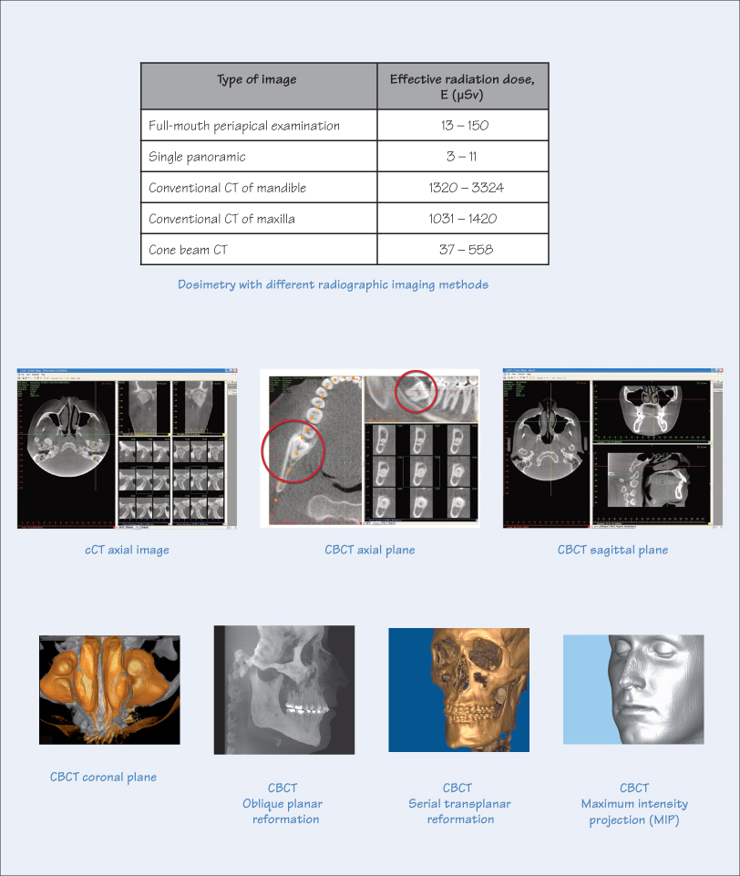
Computed tomography (CT) is a radiology-based imaging system for evaluating skeletal structures of the body. CT imaging is one method used for scanning the maxillofacial region. However, depending on the symptoms and clinical findings, alternative methods of imaging can be considered including digital radiography, magnetic resonance imaging (MRI) and ultrasonography (ultrasound imaging).
Conventional CT
Conventional CT (cCT) imaging uses a rotating helical fan beam X-ray unit and detector to scan the patient’s maxillofacial region. The ensuing digital slice-by-slice images, usually in the axial plane, are combined to create a 2-D representation. The scan can either be a single-slice or multiple detector CT (MDCT), which substantially reduces the radiation dosage. CT scans are usually limited to hospitals or specialist centres due to their prohibitive cost, large size, and the requirements for periodic maintenance, training of operating staff and adherence to strict radiation controls.
Cone Beam CT
Unlike the layered (or slice) images produced with cCT scans, cone beam CT (CBCT) scans are based on volumetric tomography. The 3-D cone beam X-ray source makes a 360° scan of the patient’s head, which is held static with an appropriate holder. The digital information obtained is in the form of 3-D cuboid blocks known as voxels or volume pixels (similar to pixels in a digital camera), representing the intensity of X-ray absorption at a particular point on the image. At predefined intervals, ‘basis’ images are acquired (usually 300–600 per scan) and subsequently reformed with algorithms using computer software to yield 3-D images in the axial (and transaxial), sagittal and coronal orthogonal planes. This process is termed multiplanar reformation (MPR). As well as images in various planes, other types of possible MPR images include the />
Stay updated, free dental videos. Join our Telegram channel

VIDEdental - Online dental courses


