Chapter 7
Tooth Eruption and Shedding
Arthur R. Hand
Departments of Craniofacial Sciences and Cell Biology, School of Dental Medicine, University of Connecticut
Tooth eruption is the movement of a tooth from its site of development in the maxilla or mandible to its functional position in the oral cavity. The eruption of a tooth requires a complex spatial and temporal coordination of molecular and cellular events among several tissues. This is made even more complicated when one considers that during much of our childhood, we have two sets of teeth in various stages of development, eruption, and shedding (Figs. 7.1, 7.2, and 7.3). That humans have two sets of teeth, twenty primary and thirty-two permanent teeth, is necessitated by the small size of the jaws in children compared to their substantially larger size in adults. The smaller size and fewer number of primary teeth can be accommodated in a child’s jaws, whereas the larger size and number of permanent teeth provide optimum masticatory efficiency in an adult. However, throughout the vertebrate subphylum, the number, formation, and eruption of teeth is highly variable. Not all vertebrates have two sets of teeth. Most fish, amphibian, and reptilian species have multiple sets of teeth, continually replacing them throughout life. Alligators, for example, have as many as 80 teeth in their jaws at one time, and may grow more than 2,000 teeth over their lifetime. Some rodents (e.g., rats, mice, hamsters) have only one set of molars, and have continuously growing incisors; the tips of the incisors chip away as the animal gnaws and bites its food. Other rodents (e.g., guinea pigs, chinchillas) have a single set of teeth, but all of their teeth grow continuously to accommodate for the wear caused by their gritty diets. Similarly, all the teeth of rabbits grow continuously, however, they have a primary and a permanent dentition.
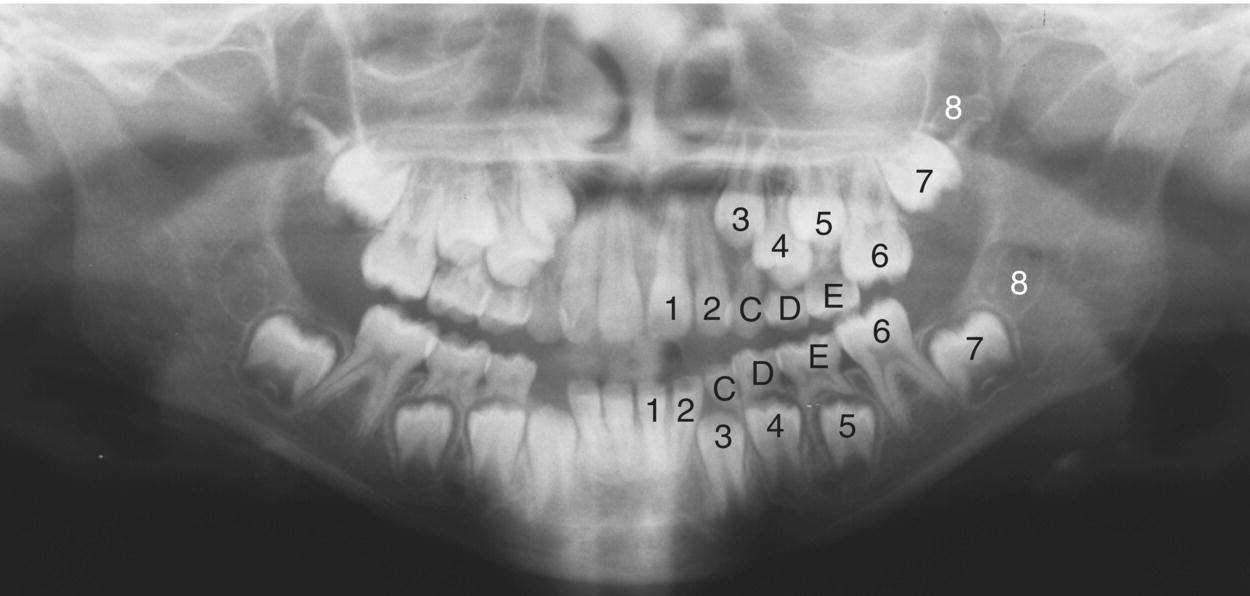
Figure 7.1 Panoramic X-ray of the mixed dentition of an 8- year-old child. The permanent teeth are indicated by the numbers (1, central incisor; 2, lateral incisor; 3, canine; 4, 1st premolar; 5, 2nd premolar; 6, 1st molar; 7, 2nd molar; 8, 3rd molar), and the remaining primary teeth by the letters (C, canine; D, 1st molar; E, 2nd molar). The four permanent incisors and the first permanent molars in each arch have erupted. The right mandibular primary canine has exfoliated, but the permanent canine has not yet erupted. The roots of the remaining primary canines and molars are undergoing resorption during the process of eruption of their permanent successors. The permanent premolars are developing between the roots of the primary molars. The roots of the second permanent molars are forming, and the earliest mineral deposition can be seen in the crowns of the developing third molars.
(Courtesy of Section of Oral and Maxillofacial Radiology, University of Connecticut School of Dental Medicine.)
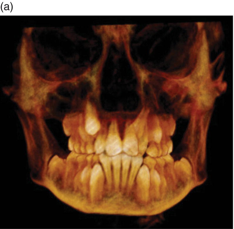
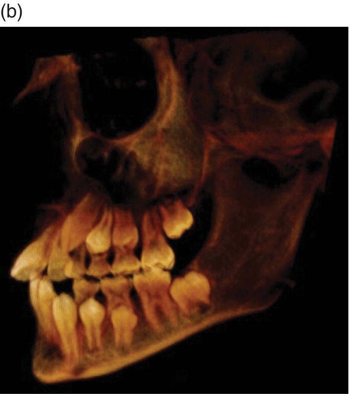
Figure 7.2 Cone beam computed tomographic (CBCT) images of the mixed dentition of an 8 year-old. (a) Frontal view. (b) Lateral view.
(Courtesy of Section of Oral and Maxillofacial Radiology, University of Connecticut School of Dental Medicine.)
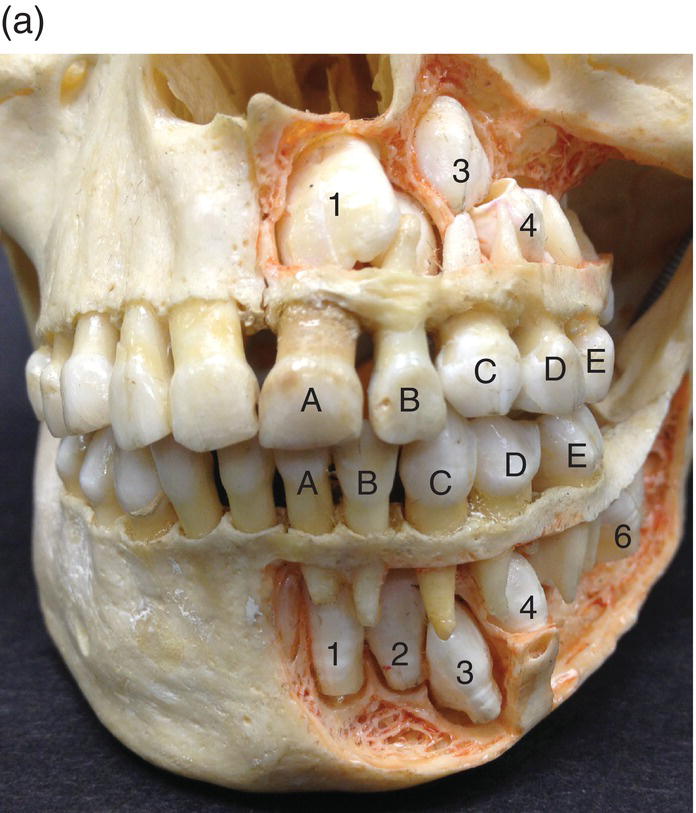
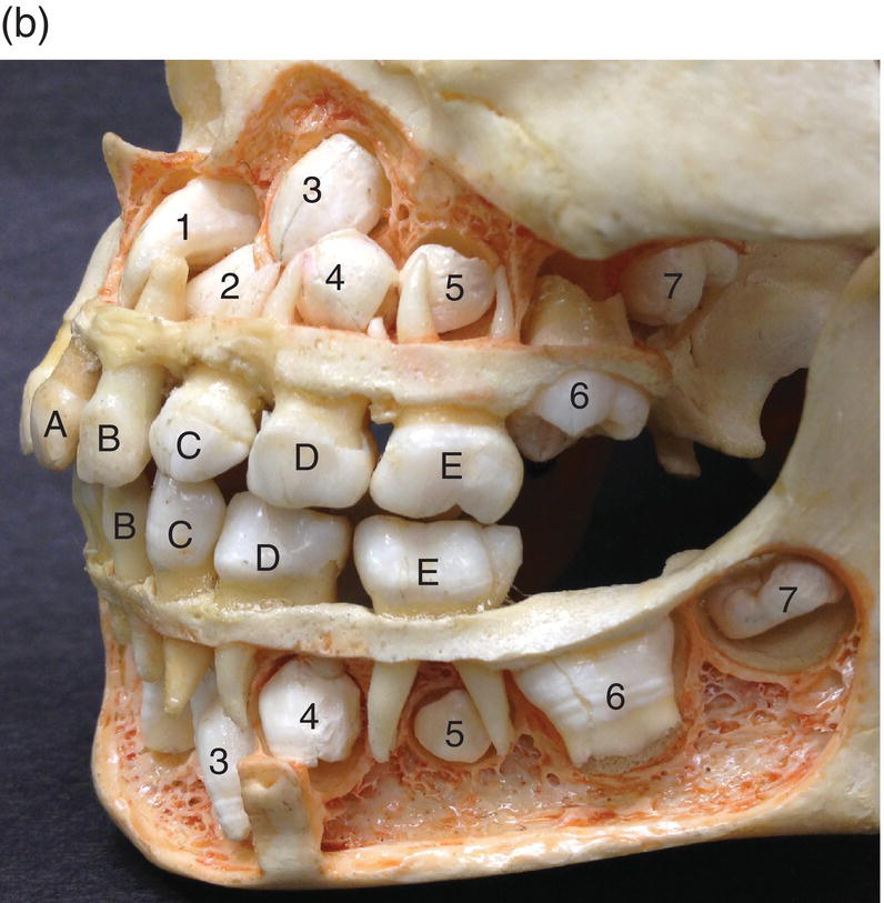
Figure 7.3 Photographs of the maxilla and mandible of a 5- to 5-1/2-year-old child with the facial cortical bone removed to reveal the developing permanent dentition. A, primary central incisor; B, primary lateral incisor; other teeth numbered as in Figure 7.1. (a) Frontal view. (b) Lateral view.
The location and timing of tooth development and eruption must be highly coordinated in order to achieve a functional and esthetic adult dentition. Eruption begins only after crown formation is complete, progresses at various speeds during different stages, and continues after occlusal contact to increase and maintain facial height throughout life and to compensate for wear of the enamel.
Normal eruption sequence
Tooth eruption occurs in a well-defined, although not invariant, temporal sequence (Tables 7.1 and 7.2). The mandibular primary central incisors are the first teeth to erupt, at about 6 months of age, followed by the lateral incisors at 7 months. The maxillary primary central and lateral incisors erupt at about 7.5 and 9 months, respectively. The mandibular and maxillary first primary molars emerge at 12 and 14 months, then the canines at 16 and 18 months, and the second primary molars at 20 and 24 months.
Table 7.1 Chronology of primary tooth formation and eruption
| Tootha | Mineralization Begins (weeks in utero) |
Enamel Complete (months postnatal) |
Emergence (months postnatal) |
Eruption Sequence | Root Complete (years) | |
| Maxillary | C. Incisor | 14 | 1.5 | 7.5 | 3 | 1.5 |
| L. Incisor | 16 | 2.5 | 9 | 4 | 2 | |
| Canine | 17 | 9 | 18 | 8 | 3.25 | |
| 1st Molar | 12.5–15.5 | 6 | 14 | 6 | 2.5 | |
| 2nd Molar | 12.5–19 | 11 | 24 | 10 | 3 | |
| Mandibular | C. Incisor | 18 | 2.5 | 6 | 1 | 1.5 |
| L. Incisor | 18 | 3 | 7 | 2 | 1.5 | |
| Canine | 20 | 9 | 16 | 7 | 3.25 | |
| 1st Molar | 12–15.5 | 5.5 | 12 | 5 | 2.25 | |
| 2nd Molar | 12.5–18 | 10 | 20 | 9 | 3 | |
aC, central; L, lateral
Table 7.2 Chronology of permanent tooth formation and eruption
| Tooth | Mineralization Beginsa | Enamel Complete (years)a |
Emergence (years)b |
Eruption Sequence | Root Complete (years)a |
||
| Maxillary | C. Incisor | 3–4 m | M 3.7 F 3.3 |
M 8.3 F 7.4 |
5 | M 10.6 F 9.3 |
|
| L. Incisor | 10–12 m | M 4.0 F 3.8 |
M 9.1 F 8.1 |
6 | M 11.1 F 9.7 |
||
| Canine | 4–5 m | M 4.9 F 4.1 |
M 11.0 F 9.4 |
8 | M 13.7 F 11.9 |
||
| 1st Premolar | 1.5–1.75 y | M 5.8 F 5.1 |
M 11.1 F 9.7 |
9 | M 13.5 F 11.8 |
||
| 2nd Premolar | 2.2–2.25 y | M 6.3 F 5.9 |
M 11.6 F 10.6 |
11 | M13.8 F 12.6 |
||
| 1st Molar | Birth | M 2.7 F 2.6 |
M 7.8 F 7.2 |
3 | M 10.1 F 9.2 |
||
| 2nd Molar | 2.5–3 y | M 6.7 F 6.3 |
M 12.4 F 11.8 |
13 | M 14.6 F 13.6 |
||
| 3rd Molar | 7–9 y | M 13.3 F 12.8 |
M 17.4 F 17.8 |
16 | M 18.2 F 18.8 |
||
| Mandibular | C. Incisor | 3–4 m | M 3.6 F 3.3 |
M 7.3 F 6.7 |
1 | M 9.2 F 8.1 |
|
| L. Incisor | 3–4 m | M 4.0 F 3.7 |
M 8.1 F 7.3 |
4 | M 9.9 F 8.8 |
||
| Canine | 4–5 m | M 4.8 F 4.1 |
M 10.9 F 9.2 |
7 | M 13.5 F 11.4 |
||
| 1st Premolar | 1.75–2 y | M 5.6 F 5.0 |
M 11.2 F 9.9 |
10 | M 13.3 F 11.9 |
||
| 2nd Premolar | 2.25–2.5 y | M 6.3 F 5.9 |
M 11.9 F 10.6 |
12 | M 14.0 F 12.8 |
||
| 1st Molar | Birth | M 2.7 F 2.6 |
M 7.8 F 7.2 |
2 | M 10.0 F 9.2 |
||
| 2nd Molar | 2.5–3 y | M 6.7 F 6.3 |
M 12.5 F 11.8 |
14 | M 14.8 F 13.8 |
||
| 3rd Molar | 8–10 y | M 13.3 F 12.8 |
M 17.4 F 17.7 |
15 | M 18.5 F 18.3 |
||
am, months; y, years
bM, male; F, female
The permanent teeth erupt in a slightly different sequence than the primary teeth. Except for the third permanent molars, the same tooth erupts a few months to more than a year earlier in females than in males. The mandibular central incisors are the first teeth to emerge, at about 6-1/2 years (in females), followed by the mandibular and maxillary first molars at about 7 years. The mandibular lateral and maxillary central incisors emerge shortly after that, then the maxillary lateral incisors at about 8 years of age. Mandibular and maxillary canines are next, at a little after 9 years, then the maxillary and mandibular first premolars between 9-1/2 to 10 years. Both second premolars emerge at about 10-1/2 years. Thus, between approximately age 6-1/2 to 11 years, a mixed dentition of both primary and permanent teeth exists. The second molars emerge at about 12 years. Eruption of the third molars is quite variable, but on average, occurs at about 17-1/2 to 18 years of age.
Histology of tooth eruption
Pre-eruptive tooth movement
The teeth develop within bony crypts of the mandible and maxilla, surrounded by the dental follicles, and initially connected to the oral epithelium via the dental lamina. The permanent successors to the primary teeth initially are located in the same bony crypt as the primary tooth, lingual (palatal) to the primary tooth (Fig. 7.4). During eruption of the primary tooth, the permanent tooth becomes enclosed in its own bony crypt. While the crown is forming the tooth undergoes small, pre-eruptive, gyratory movements. The cause and purpose of these movements are unknown, but may be related to the growth of the jaws. As the jaws increase in size, additional pre-eruptive movements serve to position the forming permanent premolar and molar tooth germs in optimal positions for their subsequent eruption. The premolar tooth germs become positioned between the roots of the primary molars; the maxillary molar tooth germs, whose occlusal surfaces originally face in a distal direction, become more vertical; and the mandibular molar tooth germs, which initially are tipped mesially, also assume an upright orientation.
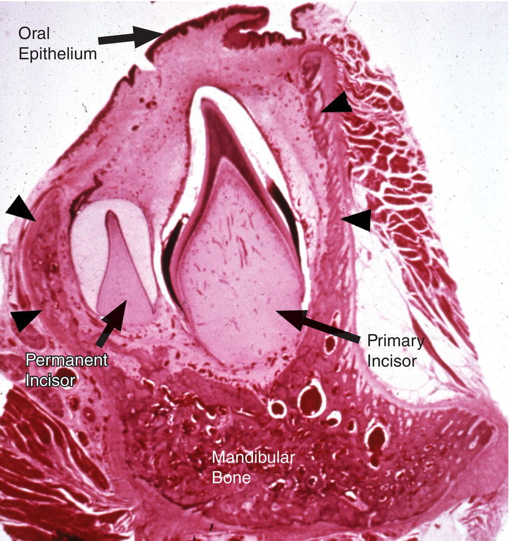
Figure 7.4 The unerupted primary and permanent incisors are developing in the same bony crypt (arrowheads) of the mandible. The late bell stage permanent incisor is located on the lingual side of the primary incisor. Some enamel matrix remains in the cervical region of the crown of the primary incisor, and its root has begun to form. As the root elongates and the tooth continues its eruption, a bony septum will form between the root and the developing permanent tooth, providing an attachment site for periodontal ligament fibers and enclosing the permanent tooth in its own crypt.
(From Moss-Salentijn, L. (1972) Orofacial Histology and Embryology: A Visual Integration. Reproduced by permission of F.A. Davis Company, Philadelphia, PA.)
Eruptive tooth movement
Intraosseous phase
Upon completion of the crown, the initial growth of the root causes some resorption of the basal bone. Subsequently, resorption of bone occurs coronally to create an eruption pathway. The rate of eruption during the intraosseous phase is slow, between 1 and 10 μm/day. Elongation of the root occurs as the tooth erupts (Fig. 7.4). If root formation does not keep pace with eruption, bone is formed apically. The main sites of bone deposition are in the interradicular areas and at the alveolar crest, the latter increasing the height of the alveolar process.
Supraosseous phase
Once the crown reaches the alveolar crest, the rate of eruption increases, up to about 75 μm per day. Most of the root growth occurs during supraosseous phase, and completion of the root also requires resorption of basal bone. Eruption to the occlusal plane requires continued root growth along with bone formation apically and at the alveolar crest and interradicular septum. However, root growth slows as the apical foramen narrows, and is very slow thereafter.
The speed of eruption is closely coordinated with bone resorption and formation. During the intraosseous phase, the rate of coronal bone resorption determines the eruption speed. During the supraosseous phase, the rate of apical bone formation and the rate of root elongation determine eruption speed. The periodontal ligament also may have a role in the supraosseous phase of eruption. The periodontal ligament does not form and the root is not attached to alveolar bone until the tooth is in the supraosseous phase.
Prior to emergence into the oral cavity, the reduced enamel epithelium covering the crown of the tooth and the basal layer of the oral epithelium undergo localized proliferation (Figs. 7.5 and 7.6). The two epithelia fuse, and the central region of the fused epithelia degenerates, creating an epithelium-lined pathway. In some cases a thin, nonvital membrane, the enamel cuticle, remains on the enamel surface, but is rapidly worn away after the tooth emerges into the oral cavity.
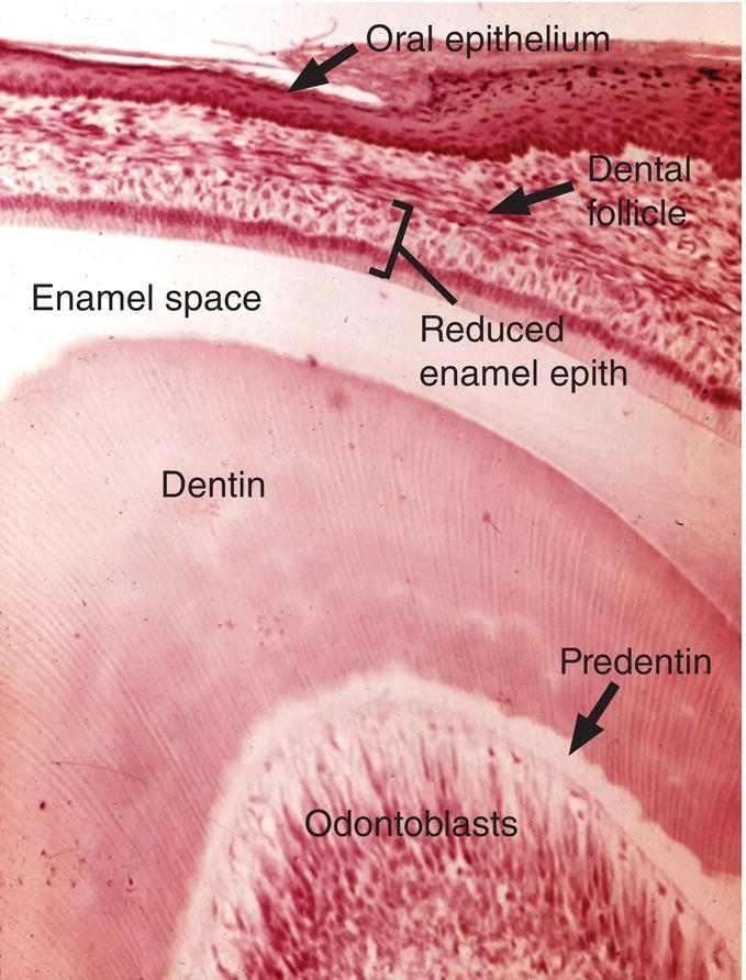
Figure 7.5 The enamel (space) of the incisor is covered by the reduced enamel epithelium and the dental follicle. A layer of columnar-shaped reduced ameloblasts is immediately adjacent to the enamel (space). The reduced enamel epithelium will fuse with the oral epithelium to create an opening through which the tooth will erupt.
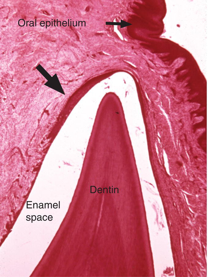
Figure 7.6 The reduced enamel epithelium near the cusp tip has proliferated and thickened (arrow) in advance of fusion with the oral epithelium.
(From Moss-Salentijn, L. (1972) Orofacial Histology and Embryology: A Visual Integration. Reproduced by permission of F.A. Davis Company, Philadelphia, PA.)
Posteruptive tooth movement
Root growth continues after emergence into the oral cavity and is not complete until the apical foramen has narrowed (Figs. 7.7 and 7.8). Eruption slows considerably as the tooth nears the occlusal plane. Slow eruption continues throughout life as the tooth functions in contact with its neighbors and the opposing teeth. This small movement allows the tooth and the periodontium to adapt to the functional demands of occlusion, to compensate for wear of the crown and to maintain facial height.
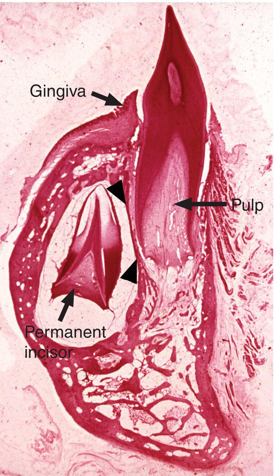
Figure 7.7 The erupted primary incisor has reached the occlusal plane, but its root is still elongating and the pulp canal is very wide. The permanent incisor is enclosed within its own bony crypt, with a thin septum of bone (arrowheads) separating it from the root of the primary tooth. The enamel of the permanent incisor is undergoing maturation, and its root is just beginning to form.
(From Moss-Salentijn, L. (1972) Orofacial Histology and Embryology: A Visual Integration . Reproduced by permission of F.A. Davis Company, Philadelphia, PA.)


