Chapter 7
Oral Pigmentation
Aim
The aim of this section is to describe the various disorders that can give rise to pigmentation of the oral mucosa.
Outcome
After reading this chapter you should have an understanding of the various conditions that can produce pigmentation of the oral mucosa, their significance, investigation and management.
Introduction
Oral pigmentation can occur as a result of deposition of exogenous pigmented substances into or on the surface of the mucosa. Alternatively it may occur as a result of deposition into the tissues of pigments produced by the body (endogenous). These latter pigments are usually melanin but may also include blood pigments such as haemosiderin.
Superficial Staining
This may be caused by a variety of food colourings, medicaments or tobacco products, and its only significance is as an indicator of the usage of such products.
Black Hairy Tongue (Fig 7-1)
This condition arises as a result of elongation of the filiform papillae and subsequent discoloration from chromogenic bacteria. The exact cause is unknown, although there is a suggestion that it may be more common in smokers and in full-denture wearers. There is no particular significance to this condition, although its appearance often concerns patients. Treatment usually involves improvement of oral hygiene, including gently scraping the surface of the tongue with either a custom-made tongue scraper or soft toothbrush.
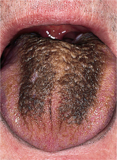
Fig 7-1 Black hairy tongue.
Amalgam Tattoo (Figs 7-2 and 7-3)
This relatively common lesion usually presents as an asymptomatic blue/black pigmented area and is often a chance finding during routine oral examination. It arises as a result of amalgam debris being implanted into the soft tissues. This subsequently breaks down releasing silver salts, which produce the staining. As a consequence, if observed over a period of time these lesions may appear to enlarge and undergo a degree of colour change. There is no specific significance to this condition, apart from its diagnosis, which could either be done by an intra-oral radiograph demonstrating the amalgam debris within the tissues, or biopsy.
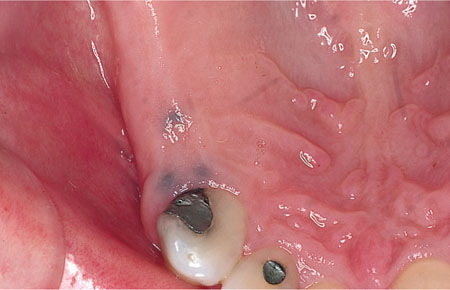
Fig 7-2 Purplish blue discoloration of the upper alveolar ridge due to implanted amalgam (amalgam tattoo).
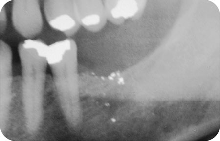
Fig 7-3 A radiograph demonstrating the presence of amalgram debris within the tissue in a patient with an amalgam tattoo.
Foreign Material
A variety of foreign substances implanted into the tissues can give rise to staining. These include graphite from pencils and deliberate tattooing. There is no specific significance to these lesions apart from their diagnosis, which may be either self-evident or through the history, although where there is doubt a biopsy will be confirmatory.
Heavy Metal Salts
Deposits of heavy metals that have been ingested, such as lead or bismuth, can be precipitated, usually as sulphides along the gingival margin producing a characteristic blue line. The significance of this is the effect on general health rather than specifically to the oral tissues.
Melanin Pigmentation
Stay updated, free dental videos. Join our Telegram channel
VIDEdental - Online dental courses


