Chapter 5
White Patches
Aim
The aim of this section is to describe those conditions that present as a white patch on the oral mucosa. Lesions that can predispose to oral cancer will be considered in the next chapter. Some conditions that can present as a white patch on the oral mucosa, such as lichen planus, are described in Chapter 2.
Outcome
After reading this section you should have an understanding of the various disorders of the oral mucosa that present as a white patch, their investigation and management.
Introduction
White patches on the oral mucosa can be broadly divided into those that can be rubbed off and those that cannot. The former comprise three main groups:
-
materia alba – this is simply the collection of debris, often food, desquamated squames and bacteria
-
Candida – for example, acute pseudomembranous candidosis
-
dead sloughing mucosa.
Those white patches that cannot be simply rubbed off are usually a result of either the development of a keratinised layer at a site that is not normally keratinised or thickening of an existing keratinised layer (hyperkeratosis). This produces a change in the texture of the tissue as well as an alteration in the optical properties, giving rise to a characteristic appearance. Although the cause and diagnosis of many of these lesions can be determined from the history or physical appearance, some have similar characteristics and, as a result, can only be differentiated histologically. For this reason, whenever there is any doubt as to the diagnosis of a white lesion, a biopsy is advocated.
Developmental White Lesions
Fordyce granules – these are clusters of creamy white spots, found mainly on the labial and buccal mucosa (Fig 5-1). They are usually asymptomatic, although when noticed by the patient may give rise to cancerophobia. Histologically they represent ectopic sebaceous glands and as such require no more specific treatment than reassurance.
Fig 5-1 Numerous ectopic sebaceous glands in the buccal sulcus (Fordyce spots).
Geographic tongue (erythema migrans) – this common condition of unknown aetiology affects between 2-4% of the population and is characterised by irregular depapilated patches, surrounded by pale, well-demarcated margins (Figs 5-2 and 5-3) . The condition predominantly affects the dorsal and lateral margins of the tongue, although similar areas can occasionally be found on other regions of the oral mucosa. The affected areas come and go over a period of days and can present at any age. The condition is usually asymptomatic, although in a few individuals discomfort can follow after eating citrus fruits or spicy foods.
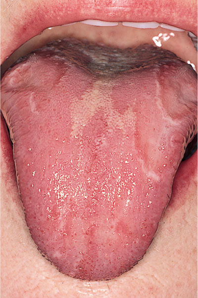
Fig 5-2 Geographic tongue (erythema migrans).
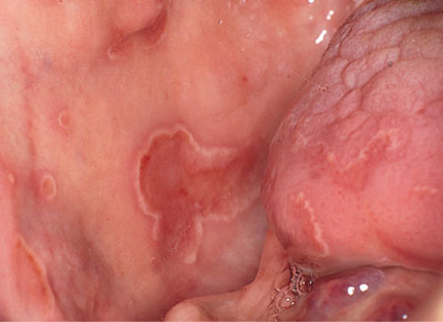
Fig 5-3 Erythema migrans affecting both the tongue and buccal mucosa. By permission of Oxford University Press from “Oral Pathology 4/e” edited by Soames, JV & Southam, JC (2005).
Geographic tongue can usually be diagnosed by its appearance alone, and biopsy is rarely indicated, unless a more sinister lesion is suspected.
The patient should be reassured about the benign nature of the condition. In those with symptoms an underlying haematinic deficiency should be excluded, as for other cases of sore tongue. Unfortunately, symptomatic geographic tongue is often unresponsive to most topical agents, including steroids. Soluble zinc sulphate tablets (45mg zinc) made into a simple mou/>
Stay updated, free dental videos. Join our Telegram channel
VIDEdental - Online dental courses


