Chapter 7
Localised Gingival Ulceration
Aim
The aim of this chapter is to detail those clinical entities that may present as a single or isolated discrete area of ulceration on the gingiva, as opposed to more widespread or multiple areas of ulceration.
Outcome
The reader should be able to differentiate between the sinister and the trivial, request relevant investigations and appropriately manage the conditions discussed.
Definition
Gingival ulceration describes an area of mucosa devoid of its surface epithelium, and exposing the underlying connective tissue.
The term ‘erosion’ is sometimes used to describe those areas of shallow ulceration that do not expose the underlying connective tissue. Table 7-1 provides a summary of the contents of this chapter.
| Major Categories | Sub Categories | Frequency of Condition | Management Setting |
| Trauma | Iatrogenic (from dental appliances) Chemical / Thermal Gingivitis artefacta |
Traumatic ulceration of the gingivae occurs frequently. | These conditions can usually be managed by non-specialists although if psychiatric morbidity is associated with gingivitis artefacta, appropriate referral is necessary. |
| Recurrent aphthous stomatitis | Minor aphthous ulceration Major aphthous ulceration Herpetiform aphthous ulceration |
Aphthous stomatitis is a very common condition (reportedly affecting 20% of the UK population). | Gingival involvement is however unusual. Initial management is undertaken in primary care. If there is a suggestion of underlying disease or poor response to treatment, the patient should be referred for a specialist opinion. |
| Neoplasia | Usually squamous cell carcinoma Lymphomas Leukaemias Metastatic tumours Benign tumours |
Oral mucosal neoplasia is uncommon and gingival involvement is rare. | Urgent referral for specialist management of malignancy. |
| Bacterial infections | Necrotising ulcerative gingivitis | Uncommon | Manage in primary care Refer if patient’s immune status is suspect. |
| Tuberculosis | Very rare | Specialist referral | |
| Syphilis | Very rare | ||
| Viral infections | Hand, foot and mouth | Uncommon | Supportive management within primary care setting |
| Varicella zoster | Uncommonly affects the gingivae | Primary care or referral dependent on distribution and severity | |
| Cytomegalovirus | Very rare | Referral – condition indicative of compromised immunity | |
| Deep mycoses | Histoplasmosis | Very rare in the UK | Specialist referral |
Traumatic Ulceration
Traumatic ulceration of the gingiva has a variety of causes including:
-
Iatrogenic, (ill-fitting or ill-designed oral appliances and inappropriate oral hygiene practices).
-
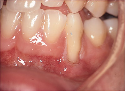
Self-induced (gingivitis artefacta) – (Fig 7-1) see Chapter 9.
-
Direct chemical toxicity (dentifrices, aspirin or the use of recreational drugs such as cocaine) – see Chapters 3 and 10.

Fig 7-1 Gingivitis artefacta.
Clinical appearance
-
Traumatic ulceration varies in appearance dependent on the causative factors. The site of ulceration may identify the source of the trauma, for example a clasp on a denture, a spring on an orthodontic appliance or malpositioned teeth.
-
The appearance is one of non-specific ulceration that may mirror very closely the source of the trauma.
-
Self-induced trauma can have dramatic clinical consequences including exfoliation of the teeth.
Clinical symptoms
-
Patients may be asymptomatic but are more likely to complain of soreness at the area of the ulceration.
-
In gingivitis artefacta the patient may well be a young adult or adolescent.
Involvement of non-gingival sites
-
Localised ulceration of the gingivae is not usually associated with extra-gingival involvement although direct chemical toxicity and physical trauma from dental prostheses or orthodontic appliances may also provoke similar manifestations on the oral mucosa.
-
Patients who self-harm may also traumatise themselves elsewhere.
Differential diagnosis
-
Non-specific ulceration.
-
Vesiculobullous disease.
-
Tumours.
-
Wegener’s granulomatosis.
-
Pyostomatitis vegetans.
-
Stewart’s midline granuloma.
Clinical investigation
-
Clinical history and examination is essential and will reveal obvious local sources of trauma.
-
Assessment of the psychological demeanour of the patient is paramount when self-harm is suspected. Direct questioning may well not elicit a truthful response, patients often strongly denying such activity.
-
Biopsy of lesions is useful to exclude other diagnoses.
Management options
-
Elimination of obvious sources of trauma.
-
In cases of self-harm, urgent referral for psychiatric advice is appropriate.
-
In cases of suspected self-harm where there is significant injury, admission to hospital for supervision may be necessary to establish a definitive diagnosis.
Bacterial Infections
Bacterial infections producing discrete localised areas of ulceration of the gingivae are uncommon.
Necrotising Ulcerative Gingivitis (NUG)
See also Chapter 9, Localised Gingival Recession.
Clinical appearance
-
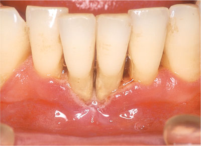
Ragged ulceration and necrosis involving the interdental papillae (Fig 7-2).
-
Lesional tissue covered by a fibrinopurulent grey slough.
-
Gingival bleeding and inflammation.

Fig 7-2 Ulceration of the interdental papillae in necrotising ulcerative gingivitis.
Clinical symptoms
-
The lesions are painful.
-
Characteristic foetor oris (bad breath).
-
Bad taste in mouth.
Aetiology
-
Anaerobic infection with a variety of organisms including Treponema vincentii and Fusobacterium nucleatum constituting the so called ‘fusospirachaetal complex’. In addition Prevotella intermedia is also reported to be associated with NUG. The risk factors that predispose to NUG include:
-
Poor oral hygiene.
-
Smoking.
-
Immunodeficiency.
-
Malnutrition.
-
Concurrent infections.
-
Involvement of non-gingival sites
-
The infection can extend to adjacent tissues, including other periodontal tissues and the oral mucosa.
-
In severe cases, in the debilitated or immunocompromised, the condition may involve the skin and can be extremely destructive (cancrum oris).
Differential diagnosis
-
Myeloproliferative disease.
-
Immunocompromised host.
Clinical investigation
-
The diagnosis is usually made on the clinical features.
-
Laboratory identification of the fusospirochaetal complex.
Management options
-
Oral hygiene instruction.
-
Smoking cessation.
-
Scaling and root surface debridement.
-
Correction of underlying predisposing factors such as malnutrition.
-
Oral metronidazole 200–400mg three times daily for three days.
Tuberculosis
Although cases of tuberculosis are increasing in incidence, oral and particularly gingival manifestations remain infrequent.
The infection may manifest on the gingivae as a solitary ulcer with irregular and often undermined margins. It may also be painless and usually results from secondary infection due to expectorated infected sputum.
Syphilis
The prevalence of sexually transmitted infections has increased dramatically over the last decade. Reported cases of syphilis in the UK are now at their highest level since 1984, with a six-fold rise in males occurring since 1998.
The primary site of infection with Treponema pallidum produces the so-called ‘chancre’ – this occurs at a variable time after inoculation, but usually of the order of three weeks later. The initial lesion is of a papule that ulcerates. The resultant chancre is indurated, often painless, associated with regional lymphadenopathy and resolves spontaneously within two to three weeks. (Alam, 2000). Gingival involvement is rare, intra-oral lesions being more frequently seen on the lips, tongue and palatal mucosa.
The superficial erosions or mucous patches (‘snail track ulcers’) of secondary syphilis are a rare gingival finding although intra-oral lesions may occur in up to 40% of cas/>
Stay updated, free dental videos. Join our Telegram channel

VIDEdental - Online dental courses


