7: Biocompatibility

Biocompatibility

Key Terms

Biocompatibility—(1) General definition: The ability of a biomaterial to perform its desired function with respect to a medical (or dental) therapy, without eliciting any undesirable local or systemic effects in the recipient or beneficiary of that therapy, but generating the most appropriate beneficial cellular or tissue response in that specific situation, and optimizing the clinically relevant performance of that therapy (Williams, 2008); (2) Long-term implantable device: Ability of the device to perform its intended function, with the desired degree of incorporation in the host, without eliciting an undesirable local or systemic effect in that host (Willliams, 2008); (3) Scaffold material for tissue engineering product: Ability to perform as a substrate that will support the appropriate cellular activity, including the facilitation of molecular and mechanical signaling systems, in order to optimize tissue regeneration, without eliciting any undesirable local or systemic responses in the eventual host (Williams, 2008).

This chapter describes the conceptual aspects of biocompatibility with specific emphasis on the solid and liquid materials of greatest relevance to dentistry. Details on test methods for biocompatibility and the monitoring of inorganic and organic species are not included because these tests are not the responsibility of practicing dentists. Many excellent textbooks and reference sources are available that describe these test methods in great detail.

Placement of a material in the body creates an interface that must exhibit both biological and structural stability during the lifetime of the implanted device. These interfaces are dynamic, and their transitional functionality is dependent on the quality of the junction and the biocompatibility of the material. The dynamics of the interfacial interaction affect the material’s biocompatibility and its acceptance by the body, which depend on the shape, size, and location of the material, its physical properties, its composition, and the stresses that develop during function.

Three major factors are linked to the success of dental materials: (1) material properties, (2) the design of the dental device, and (3) the biocompatibility of component materials. The biocompatibility of dental restorative materials is evaluated using compositional analysis, surface degradation tests, cell culture tests, clinical testing in humans, and animal model tests. The biocompatibility of a material depends on several factors:

The primary purpose of biocompatibility tests is to protect dental patients who will be treated with the materials and the office staff and lab technicians who will be handling these materials. Since no dental biomaterial is absolutely free from the potential risk of adverse reactions, the testing of biocompatibility is related to risk assessment. Thus, the challenge for the users of dental biomaterials is to select those products for which the known benefits far outweigh the known risks. Specific tests have been developed to screen restorative and implant materials for their biocompatibility. For materials, models have been developed to analyze the uptake, distribution, biotransformation, and excretion of metal ions or metal complexes in the body.

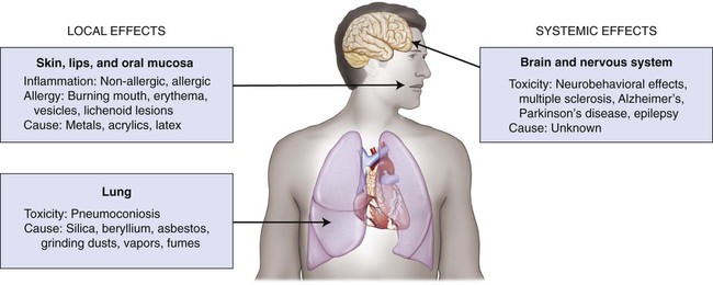
A critical adverse effect is the first event that is observed at the lowest exposure level. The location of this effect is called the critical tissue, or critical organ, and the concentration of a substance that produces this effect is the critical concentration. A metal such as mercury can be exposed to tissues as a solid binary phase (Ag2Hg3), as dissolved ions in saliva, and as atoms in the vapor form. For this metal, one must be concerned about exposure levels, absorbed dose, body burden, and critical target-tissue concentrations. Shown schematically in Figure 7-1 are the critical tissue and organ sites that can be affected by exposure to dental restorative materials and auxiliary materials used to make impressions and models. Material components can be released during melting and casting of metals, fabrication of prostheses, grinding and polishing procedures, adhesive bonding, or cementing to prepared teeth. Exposure by deposition and possibly by local uptake occurs when any one of the four types of epithelial surfaces—oral tissue, gastrointestinal tract tissue, respiratory tract tissue, and skin—makes contact with one of these metal forms.

Determining the biocompatibility of dental materials is an extremely complex task that requires consideration of cell biology, patient risk factors, clinical experience, and materials science. This chapter discusses the relevant terminology, types of biological responses that materials may cause, types of tests used to characterize biocompatibility, and anatomical aspects of the oral cavity that influence biological responses to materials. Finally, recommendations are proposed on how clinical judgments should be made in assessing the biological safety of restorative materials.

Adverse reactions to dental restorative materials and auxiliary materials include one or more of the following effects: allergic reaction, chemical burn, pulp irritation, pulp damage, thermal injury, tissue irritation, and toxic reaction. The specific causes of these effects are difficult to diagnose because of the multifactorial nature of dental treatment and the subjective nature of patients’ complaints or descriptions of their symptoms. Furthermore, there are no perfect tests for the confirmation or validation of diagnoses. For example, a visual sign of erythema in soft tissue adjacent to a new restoration may have resulted from mechanical trauma or an allergic reaction. However, an observational period may be required to determine if the effect is time limited, such as may have been caused by mechanical trauma or chemical irritation. If it is not time limited, one can assume that the erythema may have been caused by an allergic reaction. A patch test with dental test substances administered by an allergist may provide additional evidence of a potential allergy to one of the components of the material. Yet, the results of such testing are not 100% conclusive. There are no direct tests for the diagnosis of toxicity reactions. Specific tests such as urinary mercury concentration can help clinicians complement their differential diagnoses. Toxicity diagnoses are also based on the signs and symptoms presented by the patient and by a review of the history of possible exposures to a suspected toxin.

Traditionally, we have accepted the view that toxicity is dose dependent and allergy is dose independent. However, there is considerable uncertainty about which types of exposures lead to the sensitization of individuals to substances or ions released from dental restorative materials and auxiliary dental materials. Recent research suggests that dose effects for food allergies may be meaningful in describing the severity of allergic reactions. One of the common descriptors used is the dose corresponding to a 10% increase in an adverse effect relative to the control response. This is designated by ED10. An analysis of a database for 450 subjects with peanut allergy indicated that the ED10 was 12.3 mg of whole peanut (average mass of 1000 mg per whole peanut) compared with a 5% increase (ED05), corresponding to an ingested mass of 5.2 mg (Taylor et al., 2012). Thus, it is clear that sufficient data exist to establish a threshold level for a peanut allergy. However, there are no similar types of data for comparative evaluations of dose effects on the severity of allergic reactions to restorative materials. Instead, we must rely on the signs and symptoms expressed by our patients and the visual signs observed during clinical exams. In certain cases, allergy tests may have to be performed by an allergist or dermatologist (if appropriate) to assist in the diagnostic process. Specific dental test substances have been established for this purpose, and the treating dentist may have to make additional suggestions to the allergist for tests of other dental substances that are of potential significance in each case (Gawkrodger, 2005; Khamaysi et al., 2006).

This chapter is not intended to offer a course on biocompatibility test methods or theory. It is an overview of biocompatibility concepts, terminology, outcome data from national registries of adverse effects, and principles of established test methods. This overview is designed to provide a brief review of established concepts to assist dentists and other health care professionals in the conservative selection and use of dental restoratives and auxiliary materials. It also provides the essential background information and statistics on adverse events that may be needed to draft evidence-based statements on the positive and negative aspects of biomaterials and to ensure that optimal informed consent procedures on material use are employed.

Historical Perspective

Since ancient times, a wide variety of relatively inert materials have been placed or implanted in humans to replace or repair missing, damaged, or defective body tissues. Bone, seashells, animal teeth, human teeth, metals, resin materials, inorganic compounds, and other tooth replacement materials have been used for replacement of missing teeth. For the restoration of damaged or decayed teeth, metals and nonmetals have also been used, with outcomes that have varied from short-term failure to limited success in certain individuals. Many of these treatments reflected situations in which the risks were far greater than the anticipated benefits. Some of these materials have caused immediate or delayed adverse reactions because of their allergenic or toxic potentials.

Tests for the safety of restorative dental materials must ensure that a candidate material is nontoxic and unlikely to cause adverse immunological effects. Evaluations of toxicity are designed to identify adverse health events caused by physical agents, chemical agents, or both. Paracelsus (1493−1541) correctly proposed that only the dose of a substance differentiates a toxic agent from a remedy (Siddiqui et al., 2003). No pharmacological agent is free of potential toxic effects, and no drug is free from the possibility of causing an adverse event in certain individuals. No test can produce results that can guarantee that a substance will not cause adverse effects in all individuals who are treated with the substance. The allowable percentage of adverse effects in a population is based on the risks to the health and life expectancy of the individuals who will be exposed to the product under the indicated conditions and the corresponding exposure doses for its components.

Biological testing of materials has evolved significantly over the past 50 years. Since the 1980s, testing has focused on primary tests for cytotoxicity, hemolysis, Styles’ cell transformation, the Ames test, the dominant lethal response, oral LD50, intraperitoneal (IP) LD50, and the acute inhalation test. Secondary tests are also used. These include the mucous membrane irritation test (in hamsters’ cheek pouches), dermal toxicity from repeated exposures, responses to subcutaneous implantation (e.g., in rats), and sensitization (of guinea pigs). Testing of dental materials also includes tests for pulp irritation responses, pulp capping effects, endodontic applications, and dental implant performance.

As cell-culture techniques evolve, research will continue to focus on mechanisms that control the biological responses to materials. Molecular biology and imaging techniques have recently been introduced. Biocompatibility testing in the future may lead to more reliable predictions of adverse effects, and this knowledge of biological properties may allow us to formulate materials that provide specific, desired biological responses.

Influence of the American Dental Association

Methods and standards for testing the safety and effectiveness of dental materials have evolved slowly during the twentieth century. In 1930, the American Dental Association (ADA) formed a Council on Dental Therapeutics to oversee the evaluation of dental products. Also in 1930, the Council established the ADA’s Seal of Acceptance program to promote the safety and effectiveness of dental products. However, in 2005, the ADA decided to phase out the Seal of Acceptance program for professional products. Instead, a decision was made to publish a product evaluation newsletter for ADA member dentists that focused on a specific category of professional products in each article. This newsletter, called the Professional Product Review, was initiated in July 2006 and the final phase-out of the ADA seal for professional products occurred on December 31, 2007. One of the major accomplishments of the ADA was the development and acceptance of ANSI/ADA Specification No. 41. Recommended Standard Practices for Biological Evaluation of Dental Materials. This specification represented great progress toward the establishment of biological tests for dental materials.

Requirements of the U.S. Food and Drug Administration

The Dental Products Panel of the U.S. Food and Drug Administration (FDA) Medical Devices Advisory Committee reviews and evaluates data concerning the safety, effectiveness, and regulation of products for use in dentistry, and bone physiology relative to the oral–maxillofacial complex and makes appropriate recommendations to the FDA Commissioner.

In 1938, the U.S. Federal Food, Drug, and Cosmetic Act (FFDCA) authorized the FDA to oversee the safety of foods, drugs, and cosmetics. This act required evidence of drug safety before pharmaceutical products could be distributed to the public. More recently, in 1976, Medical Device Amendments (MDA) to the FFDCA included regulation of medical devices, including dental devices, for the first time. The MDA of 1976 required that FDA classify all medical devices into one of three classes, according to risk, Class I, Class II, and Class III. Dental devices, which are not specifically exempted, are required to be cleared by FDA prior to distribution into interstate commerce. Currently, the Dental Devices Branch of the Center for Devices and Radiological Health regulates premarket clearance of dental devices.

Three regulatory classes (i.e., level of control, based on risk; necessity to provide reasonable assurance of safety and effectiveness of a device type):

A brief listing of FDA device classifications and applicant requirements are summarized below.

Adverse Effects from Exposure to Dental Materials

Local and Systemic Effects of Materials

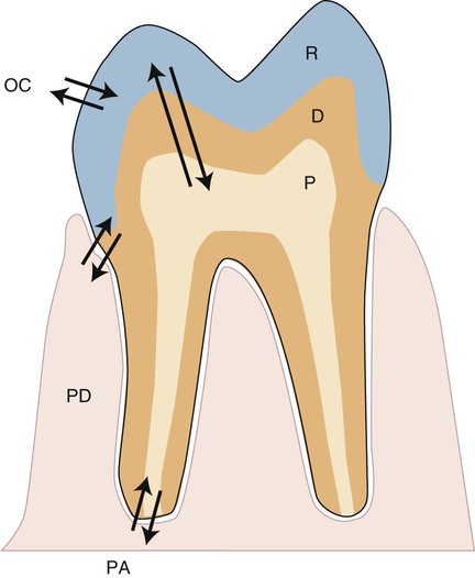
Any biomaterial that is placed adjacent to a natural tissue in the body can induce local or systemic biological effects. These effects are controlled by the substances that are released from the material and the biological responses to those substances. The nature, severity, and location of these effects are determined by the distribution of released substances. For dental materials, local effects might occur in the pulp tissue, in the periodontium, at the root apex, or in nearby oral tissues such as the buccal mucosa or tongue (Figure 7-2). The arrows in this figure indicate the pathways that foreign substances from a restorative material, if present, take into the oral environment, the tissue space next to the periodontium (PD), the pulp chamber (P), or the periapical region (PA). The periodontal ligament is also an important tissue, since it is located in proximity to the pocket or attachment area, which is often a site for accumulation of biofilms and ions, atoms, or molecules of substances released from the cervical region of dental restorations that can extend into this area. Such accumulations can be metabolized, which could then change their biological properties. These local effects are a function of (1) the ability of substances to be distributed to these sites, (2) their concentrations, and (3) exposure times, which may range from seconds to years.

In a manner similar to local effects, systemic effects from dental materials are also a function of the distribution of substances released from dental materials. Their routes of entry into the body include the following sources: (1) ingestion and absorption; (2) inhalation of vapor; (3) leakage through the tooth apex; and (4) absorption through the oral mucosa. Their migration to other sites can occur by diffusion through tissues or by flow through lymphatic channels or blood vessels. The ultimate systemic response depends on four key variables: (1) concentration of the substance; (2) time of exposure; (3) excretion rate of the substance; and (4) organ of importance or site at which exposure occurred. When substances are excreted slowly, their critical concentrations are reached more rapidly than are those concentrations of substances that are excreted quickly.

Inflammatory and Allergic Reactions

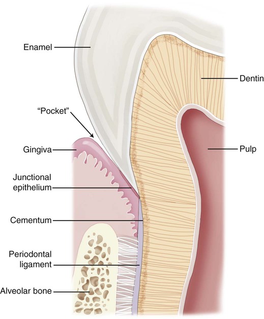
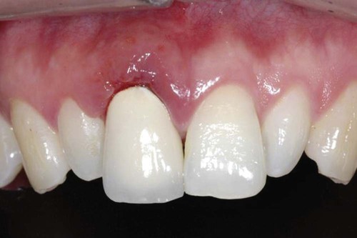
Different types of biological responses to substances can occur in humans. These include inflammatory, allergic, toxic, and mutagenic reactions. However, not all of these have been documented for dental material exposures. The inflammatory response involves the activation of the host’s immune system to ward off some challenge or threat. Inflammation may result from trauma (excessive force, laceration, and abrasion), allergy, or toxicity. Histologically, the inflammatory response is characterized by edema of the tissue caused initially by an infiltration of inflammatory cells such as neutrophils and, later in the chronic stage, to the action of monocytes and lymphocytic cells. The relationship of dental materials to inflammatory reactions is important because of chronic inflammatory responses such as pulp inflammation and periodontal disease. As indicated previously, teeth with cervical restoration margins can release ions or other substances into the gingival sulcus, and adverse reactions can affect the periodontal attachment and the periodontal ligament (Figure 7-3, with no restoration).

Allergies to substances, foods, and solid materials are well recognized by the public, but specific allergens are difficult for health care professionals to diagnose. An allergic reaction occurs when the body recognizes a substance, molecule, or ion as foreign, and the human immune system can react quickly, as during an anaphylactic reaction or slowly in delayed contact dermatitis. These reactions are insensitive to the amount of the allergen that is available or released.

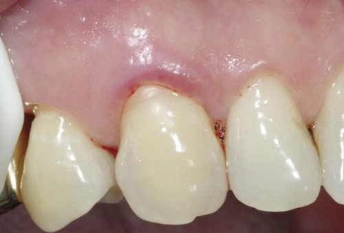
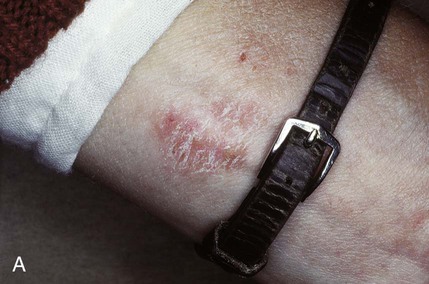
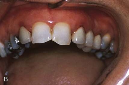
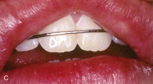
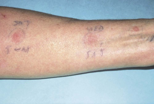
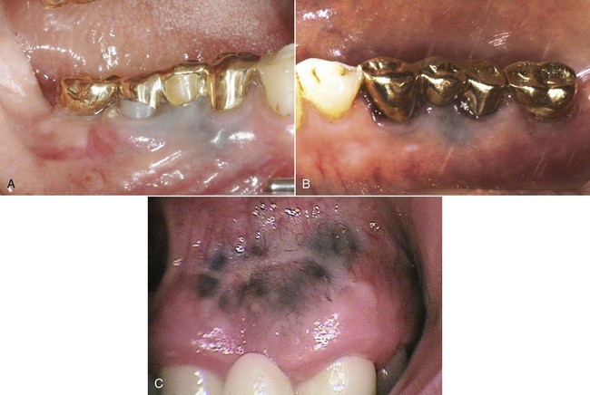
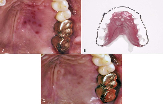
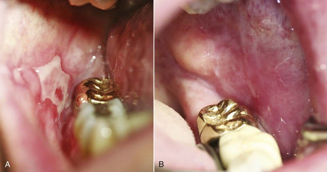
An allergic reaction induces an inflammatory response that cannot easily be distinguished from reactions caused by a nonallergic inflammatory process or by low-grade toxicity. However, by observing the signs of the effect or the absence of the signs in other locations and by the process of elimination, some reasonably logical inferences may be drawn. In some cases, observation for 2 weeks or more, when possible, can lead to a resolution of the response because the effect was caused either by trauma, another noninflammatory process, or a self-limiting allergic condition. Examples of inflammatory reactions that may be caused by allergens leached as ions from metals or other substances released by dental materials are shown in Figures 7-4 through 7-10. Figure 7-5 illustrates three potential sites for allergic reactions to nickel-containing metals: (1) a watchband buckle; (2) bilateral, fixed metal-ceramic prostheses with copings and framework made from nickel-alloy (crown on left and three-unit FDP on right); and (3) a severe reaction of lips to nickel-containing wire. Figure 7-5, D, illustrates positive responses to patch-test substances on a patient’s back. In addition, bilateral lichenoid mucositis lesions which will be discussed later, are shown in Figure 7-11 on the buccal mucosa adjacent to gold alloy crowns.

Some materials, such as latex, can cause allergy directly by activating antibodies to the material. These are classified as Type I, II, or III reactions, according to the Gell and Coombs classification of immune responses (Gell and Coombs, 1963; Rajan, 2003). These reactions occur quickly and are modulated by antibody-producing eosinophils, mast cells, or B lymphocytes. In comparison, metal ions must first interact with a host molecule to produce a delayed Type IV hypersensitivity reaction, which is modulated by monocytes and T cells. This type is often associated with contact dermatitis.

A Type I reaction (mediated by IgE or IgG4) is an immediate atopic reaction (based on a genetic predisposition to the development of immediate hypersensitivity reactions to a common environmental antigen) or anaphylactic reaction when an antigen interacts with mast cells or basophils. Atopy refers to a personal tendency, familial tendency, or both occurring in childhood or adolescence whereby one becomes sensitized and produces immunoglobulin E (IgE) antibodies in response to normal exposures to allergens, usually proteins. As a result, these individuals develop symptoms such as asthma, rhinoconjunctivitis, or eczema.

A Type II response is a cytotoxic hypersensitivity reaction, Type III is an immune complex hypersensitivity reaction, Type IV is a delayed or cell-mediated hypersensitivity, and Type V is a stimulating-antibody reaction, which is rare and sometimes classified as a subcategory of Type II (Rajan, 2002).

Precursors to Adverse Effects of Dental Materials

Each biomaterial can degrade and release components under certain environmental and physical conditions. Metals can degrade by wear, dissolution, or corrosion. The difference between dissolution and corrosion is primarily related to the difference between chemical concentration gradients and electrical current gradients. A metal restoration can release metal ions as a result of chemical reactions, electrochemical forces, or mechanical forces (such as during abrasion). For some materials, such as ceramics and resin-based composites, cyclical stresses contribute to the breakdown of the material and release of components. Thus, the biocompatibility of the material is controlled by the degradation process. The biological response to a corrosion process depends on the composition of the material, the amount of the offending species released over time, the shape of the prosthesis, and its location on or within tissues.

Corrosion is not determined only by a metal’s composition but also by the environment in contact with the metal. For example, acidic substances such as citrus juices or regurgitated hydrochloric acid alter the surfaces of ceramics. High pH environments may also increase the dissolution of some glass-phase ceramics. The environment–metal interface creates the conditions for corrosion. This interface is active and dynamic, the material breakdown products affect the body, and the body’s environment affects the material’s surface structure.

In addition to the degradation process, the biocompatibility of a material is also affected by its surface characteristics. Material surfaces are often different in composition from those of the interior structures of metals and resin-composites. Cast metals solidify first at the investment mold surface, and subsequently, the hottest area solidifies last. This transitional cooling process creates a composition gradient, which may lead to dissolution or corrosion behavior that is quite unpredictable.

Another factor that increases the potential for the release of potential allergens, mutagens, or toxins is the surface roughness of a restoration or prosthesis. For metals, a rough surface promotes corrosion, which increases the release of ions that may lead to adverse effects. Several types of beverages have caused degradation of two types of dental ceramics. Plaque accumulation also increases on roughened surfaces, and this may contribute to periodontal disease or caries.

Unfinished surfaces of resin-based composites and pit-and-fissure sealants have an oxygen-inhibited outer layer that may be more susceptible to leaching impurities such as bisphenol A. Some studies indicate that leaching of bisphenol A decreases over a relatively short period, and the results suggest that the toxicity risk is extremely low. However, the concentration of bisphenol A is dependent on the quality standards that the manufacturer follows relative to allowable impurity levels.

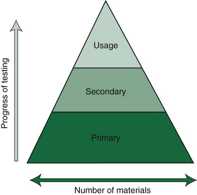
Products that pass the primary tests, such as the toxicity test, then progress to secondary and usage tests (Figure 7-12). Today, the choice of the test method is based on risk assessment, which is also divided into separate stages of analysis. Details on such testing are provided in relevant ISO standards such as ISO 14971. This risk assessment is the basis for deciding whether new tests are necessary or not, or whether data from the scientific literature are sufficient. If they are necessary, cell culture tests are regarded as the first choice combined with animal tests for sensitization. Subsequently, risk assessment continues and decisions are made on whether or not further tests (e.g., animal tests or human clinical tests) are needed. Clinical tests in some countries are mandatory for materials that are considered to be of high risk. The final decision for market clearance is then made by an interchange between manufacturers and a third party such as a governmental agency or a private organization to which this authority has been granted by a government agency. Our profession seems to be overly complacent in its acceptance of new materials without demanding proof of their safety and efficacy. When a new product is introduced on the dental market, the advertisements tend to promote the clinical performance but rarely summarize the biocompatibility tests and results of these tests compared with results from control materials.

When such products are released to the profession, dentists, dental staff, and patients must assume that sufficient safety testing has been performed to minimize potential risks. However, even with the enormous number of peer-reviewed publications that have resulted from investigations of alleged toxic and immunological reactions to mercury in dental amalgam, the evidence, thus far, has been regarded by some groups as inconclusive. However, this scenario raises a most important question: How much evidence is sufficient to demonstrate that a product is sufficiently safe for general clinical use? Dentists must rely on the appropriate test methods that are required in standards and in legal regulations and assume that sufficient evidence of safety has been established for dental devices once they have been cleared for market use.

Since toxicity is dose dependent, it is obvious that materials that release too much of a substance can cause overt toxicity. The terms and definitions given previously indicate that there are different dose thresholds for various levels and probabilities of risk. Figure 7-13 shows a plot of cellular glutathione from monocytes that were exposed to mercury or palladium ions in a cell culture. Mercury ions are shown to increase the glutathione content of the monocytes in the cell culture, while palladium ions cause the glutathione content to decrease. Since glutathione is essential for maintaining the redox balance in the cells, exposure to these metallic ions can change the cellular function of the monocytes.

Immunotoxicity

Our understanding of the differences between toxicity, inflammation, allergy, and mutagenicity has become clearer as we learn more about the interactions between biomaterials and cells. The principal concept of immunotoxicity is that substances leached from materials can alter immune system cells, resulting in enormous biological consequences because of the amplifying nature of immune cells. These cellular alterations can occur initially because of the toxic effect of a leached substance. Monocytes control chronic inflammatory and immune responses, and they also secrete many substances that alter the actions of other cells. Thus, if substances leached from a biomaterial change the monocyte’s ability to secrete these substances, the biological response can be greatly influenced and this may greatly impair cellular defense mechanisms against bacteria (Schmalz et al., 2011).

As stated earlier, cell function can either increase or decrease as a result of immunotoxic effects. As shown in Figure 7-13, mercury ions are known to increase the glutathione content of human monocytes in cell culture, whereas palladium ions decrease the cells’ glutathione content. Glutathione is important in maintaining oxidative stress in cells, and any change in its concentration can alter cell function. Higher concentrations of mercury can also decrease glutathione as the ion concentration becomes more toxic.

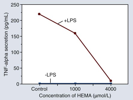
Figure 7-14 shows a graph of the amount of tumor necrosis factor alpha (TNF-α) secreted by monocytes after exposure to hydroxyethyl methacrylate (HEMA) in different concentrations. The +LPS line represents the effect of cell stimulation by lipopolysaccharide while the –LPS line indicates that the monocytes were not stimulated by lipopolysaccharide. The significance of this effect is that relatively small amounts of HEMA released from bonding adhesives or resin-based composites can alter the normal functions of monocytes, thereby contributing to the potential immunotoxicity of some resin-based products.

Pulpal and Periodontal Effects

The postoperative discomfort or pain caused by treatment with dental materials may result from any of several factors, including thermal trauma, chemical injury, microleakage, and allergy. The hydrodynamic theory of pulp pain is related to the movement of dentinal fluid and its influence on the odontoblastic processes. It is well known that dentin permeability increases substantially in areas closer to the pulp chamber. Thus, the damaging effect of a material, if such an effect occurs, is strongly influenced by the remaining thickness of dentin between the material and the pulp chamber. This outward fluid pressure from the pulp chamber toward the enamel is not sufficient to eliminate the inward diffusion of bacteria, bacterial products, or material components into the pulp. A major problem in the diagnosis or potential pulp damage caused by a material or substance is the fact that there is virtually no correlation between the histologically documented damage to the pulp and clinical symptoms. This is considered as a major drawback of clinical studies in this area.

Because the cervical margins of many dental restorations are near the periodontal attachment area (see Figure 7-3), the biocompatibility of these materials may influence the body’s ability to defend against bacteria that cause periodontal disease. Further, the periodontal pocket, or gingival sulcus, may accumulate significant concentrations of leached substances that do not accumulate to these levels in other areas. In addition to the accumulation of leached or dissolved substances in subgingival areas, substances that are leached from root canal filling materials may accumulate next to the apical foramen. Substances that accumulate in these areas can lead to inflammatory reactions, allergic reactions, periodontal pathology, and periapical lesions.

Interfaces with Dental Materials

Tooth-supported dental restorations consist of one or more prepared teeth, a monolayer or multilayer restorative material, and auxiliary dental biomaterials such as dental adhesives, cements, and sealing agents. Although several interfaces may be present in these restorations, the dentin–cement or dentin–resin interfaces are the most important in transition areas for transfer of leached substances into dentinal fluid. Dental cements such as zinc phosphate, glass ionomer, zinc polycarboxylate, and zinc oxide-eugenol do not require etching of dentin to be used as luting agents. In contrast, adhesive-bonded, resin-based cements may require acid etching of dentin to remove the smear layer and to expose the collagen mesh that allows infiltration of the bonding resin. This resin layer acts as a partial barrier to the transport of elements, ions, or substances that are released from a variety of restorative materials.

If the resin material does not penetrate the collagenous network or debonds from it as the resin shrinks during polymerization, a microscopic gap will form between the resin and dentin. This shrinkage may also occur with enamel. Although this gap is only a few microns wide, it is wide enough to permit bacteria to penetrate this interfacial space, since the average size of a Streptococcus bacterium is only about 1 µm in diameter. The dentin–resin interface occurs when the clinician attempts to bond resin-based restorative materials to dentin. The interface actually forms between the resin and the collagen network. Thus, the integrity of the resin–collagen interface will control the potential pulp-damaging effect of these restorations.

As shown in Figure 7-15, incomplete bonding or resin penetration into the collagen mesh of acid-etched dentin can lead to fluid ingress along gaps wider than 1 µm, which is referred to as microleakage. The microscopic gap for the so-called microleakage process may lead to several undesirable events. The bacteria that migrate to the pulp may initiate an infection of pulp tissue. The gap also promotes material breakdown along the unsupported margin. This breakdown increases the gap width, which allows larger particles and molecules to progress toward the pulp chamber. It also causes marginal staining and compromised esthetics that may lead to premature replacement. One in vitro study revealed less leakage for amalgam restorations compared with resin-bonded composites (Őzer et al., 2002).

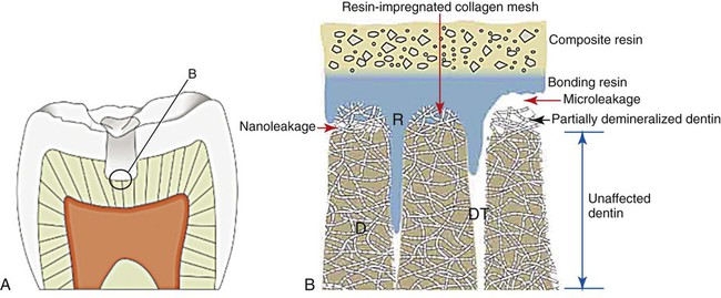
If the resin penetrates the collagen network of dentin but does not penetrate it completely, then a much smaller gap (less than 0.1 µm in most cases) will exist between the mineralized matrix of dentin and the collagen–resin hybrid layer (see Figure 7-15). This much smaller gap has been claimed to allow nanoleakage, which probably does not allow bacteria or bacterial products to penetrate the marginal gaps of the restoration and the pulp. However, fluid exchange most likely occurs, and this may degrade the resin or the collagen network that is incompletely embedded with the resin, thereby reducing the longevity of the dentin–resin bond.

Nanoleakage is not known to occur between restorations and enamel because enamel contains virtually no organic mass and therefore has no collagenous matrix into which a resin may penetrate. Although it is unclear whether leakage toward or into the dental pulp chamber is a major factor in the biological response to dental materials, one must be aware of potential immune responses in the pulp and periapical tissues that may occur independently of leakage phenomena.

Influence of Biocompatibility on the Osseointegration of Implants

The success of endosseous dental implants is based on the biocompatibility of the implant surface and the ingrowth of new bone into the surface through the process of osseointegration. Very few implant materials or implant coatings promote osseointegration. The most common implant materials include (1) CP Ti; (2) titanium-aluminum-vanadium alloy; (3) tantalum; and (4) some types of ceramics. Materials that allow osseointegration have very low degradation rates, and they tend to form surface oxides that enhance bony approximation. Some materials such as bioglass ceramics promote a perfect osseointegration of the bone. The general process of biointegration involves the adaptation of bone or other tissue to the implanted material without any intervening space along the tissue-material interface (see Chapter 20).

Mercury and Amalgam

The controversy over the biocompatibility of amalgam has waxed and waned several times in the 170-plus year history of its dental use in the United States. Most of the controversy stems from the known toxicity of mercury and the question of whether mercury from amalgam restorations has toxic effects. Mercury occurs in four forms: as the metal (Hg0), as an inorganic ion (Hg2+), as a component of the silver-mercury phase, or in one of several organic forms such as methyl or ethyl mercury. Metallic mercury gains access to the body via the skin or as a vapor through the lungs. Ingested metallic mercury is poorly absorbed from the gut (0.01%), so the primary portal into the body is through inhalation of mercury vapor.

Absorption of specific metals through the oral mucosa, gastrointestinal tract, or respiratory tract can vary considerably for different chemical forms of a metal. Excretion may occur through exhaled vapor or through urine, feces, or skin. For example, mercury vapor is readily absorbed after inhalation. Dissolved mercury can be transported through blood and distributed to the brain and other organs and excreted by exhalation and in urine. Elemental mercury is transported to blood cells and tissues, where it is oxidized rapidly to mercuric mercury (Hg2+).

The most common forms of mercury that occur naturally in the environment are metallic mercury, inorganic salts, mercuric sulfide (HgS), mercuric chloride (HgCl2), and methyl mercury (Ch3Hg+). Mercury forms numerous compounds, assuming +1 valence in mercurous compounds and +2 valence in mercuric compounds. The transformative ability of mercury can be either helpful or harmful. Microorganisms and various natural processes can convert metallic mercury to inorganic mercury compounds, inorganic mercury compounds to organic mercury compounds, and organic to inorganic compounds. Methyl mercury is the most common form that is transformed by natural processes; it is a more toxic form than ethyl mercury (C2H5Hg+) or elemental mercury. Methyl mercury is a major safety concern because it bioaccumulates through the food chain; its pharmacokinetic half-life is longer (1.5 to 3 months) than that of ethyl mercury (less than 1 week). Moreover, it is accumulated in the body rather than being excreted in the gut, as occurs with ethyl mercury. In air, the concentrations of mercury range from about 0.01 to 0.02 µg/m3.

Mercury accumulates in the kidneys. In the brain, metallic mercury can be converted to an inorganic form that is retained in the brain. Elemental mercury and mercury vapor have a half-life of 1 to 3 months. Mercury leaves the body by excretion through urine and feces.

Exposure to high levels of mercury can injure the brain, kidneys, and the developing fetus. The nervous system is sensitive to all forms of mercury, although the brain is most sensitive to metallic mercury and methyl mercury. Chronic mercury toxicity may be manifested as tremors; memory loss; and changes in personality, vision, and hearing. Children and fetuses are most sensitive to the effects of mercury on the nervous system. Selenium, an essential element, is claimed to be protective against the toxic effects of mercury.

Mercury is not regulated under the Clean Air Act (U.S. Code, Title 42, Chapter 85, signed by President Richard Nixon on December 31, 1970). The Clean Air Act is the law that defines the responsibilities of the U.S. Environmental Protection Agency for protecting and improving air quality and the ozone layer. The Clean Air Act Amendments were enacted by Congress in 1990 and legislation has made several minor changes since 1990. For air in the workplace, the Occupational Safety and Health Administration has set a permissible exposure limit (PEL) for mercury vapor in air of 0.1 mg/m3.

The United Nations Environment Programme (UNEP) has organized global meetings through the UNEP Global Mercury Partnership to protect human health and the global environment from the release of mercury and its compounds by minimizing and, where feasible, ultimately eliminating global, anthropogenic mercury releases to air, water and land. Since 2001 the Governing Council/Global Ministerial Environment Forum of UNEP has discussed the need to protect human health and the environment from the releases of mercury and its compounds. By the end of 2011, three sessions of the Intergovernmental Negotiating Committee (INC) organized by UNEP had been held to prepare a Global Legally Binding Instrument on Mercury: INC1 (June 7-11, 2010, Stockholm, Sweden), INC2 (January 24-28, 2011, Chiba. Japan), and INC3 (October 31-November 4, 2011, Nairobi, Kenya). Although no globally-binding document on controlling mercury in dental amalgam and other mercury-containing products had been approved, a proposal for a “phase-down” of dental amalgam was supported combined with a need for research on more durable alternatives to dental amalgam and a renewed emphasis on prevention of caries and the associated need to reduce the need for restorations throughout the world.

Biological monitoring of the metal species and content in urine, whole blood, plasma, or serum is recommended for specific metal species depending on the level of biological risk and the critical target tissues that are involved. Sampling of metals in hair, nails, feces, bone, and teeth is also advocated in certain cases.

Several studies have shown that amalgams release sufficient vapor to cause absorption of between 1 and 3 µg/day of mercury, depending on the number and size of amalgam restorations present (Langworth et al., 1988; Berglund, 1990; Mackert and Berglund, 1997; Ekstrand et al., 1998). The inhaled mercury gains access to the bloodstream via the alveoli of the lungs. From the blood, mercury is distributed throughout the body, with a preference for fat and nerve tissues. Mercury is also ingested as particles produced by wear, and about 45 µg/day of mercury may reach the gut either as the amalgam form or as dissolved and released Hg2+ ions. The absorption of ionic mercury is also poor (approximately 1% to 7%). Mercury trapped in amalgam particles is also poorly resorbed. Methyl mercury is not produced from amalgams but is generally a product of bacteria or other biological systems acting on metallic mercury. Methyl mercury is the most toxic form of mercury and is also very efficiently absorbed from the gut (90% to 95%). Methyl mercury is absorbed mainly from the diet, particularly from fish (especially shark, swordfish, and tuna), which contribute a significant portion.

Concerns about mercury stem from its toxicity and its relatively long half-life in the body. The toxicity of mercury is well known; the symptoms depend somewhat on the form. Acute symptoms are neurologically based or kidney based, ranging from paresthesia (at 500 µg/kg or above) to ataxia (at 1000 µg/kg or above), joint pain (at 2000 µg/kg or above), and death (at 4000 µg/kg or above). The lowest known level for any observable toxic effect is 3 µg/kg. This level translates to about 30 µg of mercury per gram of creatinine clearance in the urine. At chronic exposure levels, the symptoms are more subtle and include weakness, fatigue, anorexia, weight loss, insomnia, irritability, shyness, dizziness, and tremors in the extremities or the eyelids. Although amalgams do not release anywhere near toxic levels of mercury, the long half-life of mercury in the body raises concerns among some individuals. The half-life ranges from 20 to 90 days, depending on the form, with methyl mercury exhibiting the longest half-life and inorganic forms the shortest. Numerous tests for the body burden of mercury have been developed, including those based on the analysis of blood, urine, and hair. Of these test parameters, measurement of mercury in the urine after 24 hours may be the best long-term indicator of the total metallic mercury body burden, normalized to grams of creatinine clearance from the kidneys.

Humans are exposed to mercury from a variety of sources in addition to dental amalgams. There are extremely sensitive methods for detecting mercury in parts per trillion; these methods have made it possible to analyze the sources of mercury exposure for humans. Estimates of intake levels from air (in micrograms per day) are 0.12 for Hg0, 0.04 for Hg2+, and 0.03 for methyl mercury. Water probably contributes about 0.05 µg/day and food about 20.0 µg/day in the form of Hg2+. Depending on one’s diet, the consumption of fish contributes about 0.9 µg/day of Hg0 and 3.8 µg/day of methyl mercury. These values place the 1 to 3 µg/day of absorbed Hg0 vapor from amalgams in perspective. Thus, the intake of mercury is a complex issue with many sources and forms of exposure. Furthermore, there is considerable variability from individual to individual depending on diet, environment, and dental status. Despite the confirmed exposure of humans to these low levels of mercury, the biological effects of these levels are insignificant.

Numerous studies have attempted to determine whether mercury exposure from dental restorations or other sources contributes to any documentable health problem. Several studies have estimated the number of amalgam surfaces needed to expose an individual to mercury concentrations with a minimum observable effect (slightly impaired psychomotor performance, detectable tremor, and impaired nerve conduction velocity). Estimates are that several hundred amalgam surfaces would be necessary to achieve these levels. Even if all 32 teeth were restored on all surfaces with amalgam, the total number of surfaces (counting incisal edges) would be only 160. Other studies have measured renal function in patients in whom all of the amalgam was removed at the same time (the worst possible case). Despite markedly elevated blood, plasma, and urine levels of mercury, no renal impairment was noted. Still other studies have attempted to look at blood cell types and cell numbers in dentists, who are presumably exposed to higher levels of mercury because of their daily occupational exposure. No effects of mercury have been noted. Other studies for neurological symptoms in children populations occupationally exposed have shown no effects (Bellinger et al., 2006, 2007; DeRouen et al., 2002, 2006). In summary, there are simply no data to show that mercury released from dental amalgam is harmful to the general population.

Estrogenicity

In 1996, a research group claimed that dental sealants released estrogenic substances in sufficient quantities to warrant concern (Olea et al., 1996). Since then, the estrogenicity of dental composites has also been questioned, particularly for use in children. Estrogenicity is the ability of a chemical to act as the hormone estrogen does in the body. If these chemicals are not indigenous to the body, the substance is called a xenoestrogen. The occurrence of xenoestrogens in the environment has been a concern for many years. Environmentalists fear that these substances will alter reproductive cycles and developmental processes in wildlife, and there is evidence to support these concerns with regard to humans. The concern about estrogens in dentistry centers around a chemical called bisphenol A (BPA), which is a synthetic starting point for bis-GMA (bisphenol-A-glycidyldimethacrylate) composites in dentistry as well as many other plastics. The fear is that the release of these substances might alter normal cellular development or cell maintenance if BPA has estrogenic effects, even at the impurity levels that may be encountered in practice. It is generally accepted that BPA is released from bis-DMA, which is also used in dental products. However, if bis-DMA is used, the amount released after placement of a restorative filling is too small to be of concern. However, to overcome any concerns, products free of bis-DMA can be used.

There is fairly convincing evidence that BPA and BPA dimethacrylate may act on the estrogenic receptors in cells. Thus, these chemicals are probably xenoestrogens. This evidence is derived from mo/>

Stay updated, free dental videos. Join our Telegram channel

Jan 1, 2015 | Posted by in Dental Materials | Comments Off on 7: Biocompatibility

VIDEdental - Online dental courses

Get VIDEdental app for watching clinical videos