(b) 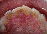
Table 7.2.1 Clinical features of lateral luxation injuries
|
Parallax view intraoral radiographs (Figures 7.2.2a and b) of the upper central incisor teeth and a limited volume cone beam computed tomography (CBCT) scan of the same area (Figure 7.2.3) were taken to discern the exact nature of the injury. The intraoral periapical radiographs demonstrated widening of the periodontal ligament space on the mesial aspect of the 11. However, there was little else of significance. The remaining maxillary anterior teeth had a normal radiographic appearance on these conventional radiographs. In contrast to the periapical radiographs, the CBCT scan clearly demonstrated the full extent of the injury to the 11. The crown of the tooth had been displaced palatally, while the root of the tooth had rotated labially, resulting in a fracture of the labial cortical plate. The tooth had consequently become engaged and locked in the alveolar bone. The tooth did not sustain any root fractures. The ‘locked’ position of the tooth explained its lack of clinical mobility and the high-pitched tone it produced when percussed. Table 7.2.2 outlines radiographic features of lateral luxation injuries.
Figure 7.2.2 (a) Pre-operative periapical radiograph (left) of the 11 shows a widening of the periodontal ligament space on the mesial aspect of the 11, (b) pre-operative periapical radiograph (right) with a shift in the X-ray tube to the right.
(a) 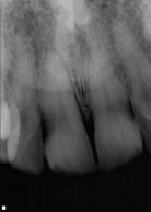 (b)
(b) 
Figure 7.2.3 A CBCT scan confirms the lateral luxation injury of the 11. (a) Axial, (b) coronal, and (c) sagittal reconstructed images clearly show the nature of the luxation injury to the 11. The labial displacement of the root has resulted in an increased palatal periodontal space (yellow arrow) and a consequent fracture of the alveolar cortical plate due to the buccal rotation of the root (red arrow). The crown of the tooth had been palatally luxated, resulting in the root being pushed labially.
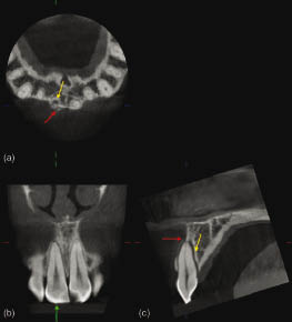
Table 7.2.2 Radiographic features of lateral luxation injuries
|
Diagnosis
What is the diagnosis?
The diagnosis is lateral (palatal) luxation of the upper right central incisor (11).
Why were two radiographs and a CBCT scan taken?
Stay updated, free dental videos. Join our Telegram channel
VIDEdental - Online dental courses


