Variations in Transition of Maxillary Posterior Teeth*
The space available in the dental arch for the transition of the maxillary posterior teeth is bounded as in the mandible by the distal surface of the crown of the lateral permanent incisor and by the mesial surface of the crown of the first permanent molar. The sum of the mesiodistal crown dimensions of the maxillary deciduous molars and canine is slightly larger than that of their successors. The first premolar has approximately the same mesiodistal crown dimension as its predecessor. The second premolar is usually smaller than the preceding deciduous molar; however, the difference between the two is less marked than between the corresponding mandibular teeth. The maxillary permanent canine has a considerably larger mesio-distal crown dimension than the deciduous one. This difference is larger for boys than for girls and more marked in the maxilla than in the mandible.71 The excess in the sum of the mesiodistal crown dimensions of the deciduous molars and canine in relation to their successors is smaller in the maxilla than the mandible. Yet the average leeway space is larger in the maxillary dental arch. Seemingly a contradiction, it is related to the occurrence of more and larger diastemata between the teeth of the involved segment of the dental arch. The disparity in the transition of the maxillary posterior teeth and that of the mandibular teeth is related not only to the differences in mesiodistal crown dimensions and in the leeway space; size and shape of the middle section of the apical area and the sequences of emergence also play a role.
The transition of the maxillary posterior teeth is discussed in relation to the space available for the permanent canine and the premolars in the middle section of the apical area and in the dental arch. Variation in the location of the permanent teeth concerned, and their relation to each other, to their predecessors, and to the adjacent lateral permanent incisor and first molar are indicated. The differences in emergence sequence and associated preemergence and postemergence behaviour are dis-cussed. Seven transition patterns are distinguished, five of which are presented in detail.
As has been described before, the anterior section of the apical area in the maxilla is bounded laterally by the permanent canines. Before their emergence, the large permanent canine crowns limit the space initially available for the forming permanent incisor crowns and subsequently for their roots. After complete eruption of the permanent canines their apices form the lateral boundaries of the anterior section of the apical area. The room then normally available is adequate to contain the relatively small roots of the permanent incisors. As explained previously, the anterior section of the apical area is initially too small to house the forming permanent crowns in an arrangement comparable to what they will occupy after their eruption is completed. A corresponding situation exists in the maxilla for the middle section of the apical area. In the sagittal direction the middle section of the apical area is too small to house the forming premolars and permanent canine at the same level. Further, the size of the transverse dimension and the presence of the deciduous teeth roots allow only a limited variation in buccopalatal direction. The first premolar and permanent canine are formed close together, and their crowns occupy positions of different levels prior to their eruption.
The size and shape of the middle section as well as of the anterior section depend upon the morphology and location of the piriform aperture. The permanent canines are formed lateral to the anterior section and are, of all teeth, at the greatest distance from the occlusal plane. The permanent canines will develop the longest roots of the entire dentition. In the preeruptive phase the premolars are distinctly closer to the occlusal plane than the permanent canine. The initial arrangement of the premolars and permanent canine in the maxilla resembles that in the mandible. However, there are marked differences. The maxillary canine is not usually situated mesial to the first premolar crown but superiorly, and it overlaps it vertically. The distal corner of the permanent canine crown develops almost in contact with the mesial cementoenamel junction of the first premolar. The mesial concavity of the first premolar can be thought to result from this relation.
In accordance with the situation in the mandible the crown tip of the maxillary permanent canine is located palatally to the apex of its predecessor. The mesial part of the crown is in close proximity to the root of the fully erupted lateral permanent incisor and frequently overlaps the root buccally. The premolars are located between the roots of their predecessors. The first premolar is more to the buccal and closer to the occlusal plane than the second premolar. Compared to the mandible, the space between the mesial surface of the first premolar crown and the distal surface of the root of the lateral permanent incisor is smaller.
The middle section of the apical area in the maxilla shows a large individual variation in size and shape. The differences appear primarily in the location of the canine and first premolar. Considerable variation, more than in the mandible, is encountered in the sagittal and transverse locations of these teeth. As in corresponding previous chapters, prepared human skulls serve to demonstrate the variation in size and shape of the middle section of the apical area and the associated arrangement of the forming permanent teeth (Figs. 6-1 to 6-3).
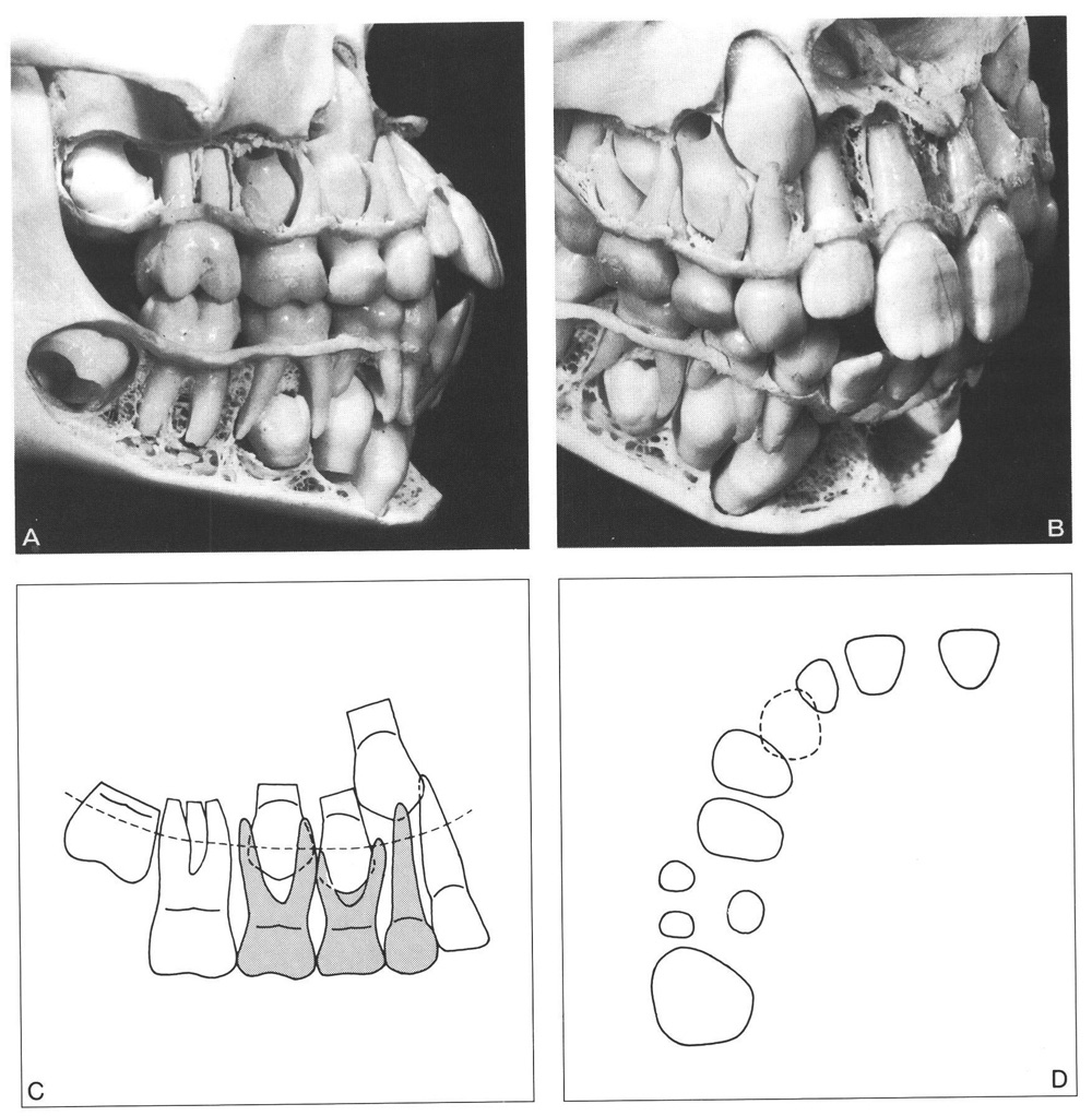
Fig. 6-1 Large middle section of the apical area in the maxilla. The relation between the mesiodistal crown dimensions of the maxillary permanent canine and the premolars and the size and shape of the middle section of the apical area is favourable. However, no diastemata are present in the dental arch.
A The permanent canine is located more superiorly and palatally than the premolars and is mesially angulated. This is in accordance with the morphology of the adjacent structures. Both maxillary premolars are closer to the occlusal plane and at about the same level. The second deciduous molar has a larger mesiodistal crown width than its successor.
B The permanent canine crown slightly overlaps the root of the lateral incisor. Its cusp tip is palatal and slightly mesial of the apex of its predecessor. The canine partly overlaps the first premolar in a mesiodistal direction. Its distal corner is in close proximity to the first premolar. The deciduous canine crown is smaller than the permanent one.
C Schematic drawing with a dotted line indicating the section presented in D. Both premolar crowns are about the same level and in a comparable relation with the roots of their predecessors. Some space exists between the mesial surface of the first permanent molar root and the second premolar crown. The first premolar crown is mesially inclined and close to the second premolar, which is normally oriented perpendicular to the occlusal plane.
D The premolar crowns are not in contact with each other and do not overlap. Both are harmoniously positioned in a buccopalatal direction. The distance between the first premolar crown and the lateral incisor root is relatively large. A t this stage of development the permanent canine crown overlaps both adjacent teeth slightly. However, later on there will be sufficient room in the alveolar process to house the root of the permanent canine with its smaller circumference.
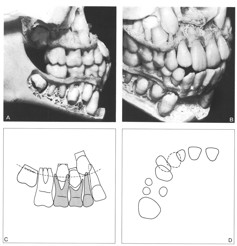
Fig. 6-2 Medium middle section of the apical area in the maxilla. The position of the permanent canine crown, its proximity to the lateral permanent incisor root and first premolar crown, and the differences in the mesiodistal crown dimensions of the corresponding deciduous and permanent teeth are critical.
A Both premolars are close together. The second premolar is situated close to the first permanent molar. The mesiodistal crown dimensions of the deciduous canine and molars are small in relation to those of their successors.
B The permanent canine is medially situated above the first premolar, almost totally overlapping it in the vertical plane. Both teeth are mesially angulated, particularly the canine. In addition, the first premolar is mesiopalatally rotated, extends buccally, and is situated closer to the occlusal plane than the second premolar. The mesial surface of the canine crown extends anteriorly to the line that can be constructed as the continuation of the mesial surface of the lateral incisor root.
C The differences in vertical position of the crowns of the premolars and permanent canine are associated with the overlapping of these teeth. The distance in the dental arch between the mesial surface of the first permanent molar and the distal surface of the lateral incisor is small.
D The second premolar is located more palatally and the first premolar more buccally than normal. The space available between the mesial surface of the first premolar and the distal surface of the lateral incisor is limited.
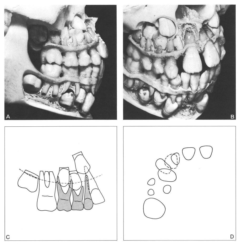
Fig. 6-3 Small middle section of the apical area in the maxilla in an instance where the lateral deciduous incisor was lost prematurely and its successor has not yet emerged.
A The permanent canine crown is in a superior position and markedly mesially angulated. The forming part of the first premolar is located buccal to the canine crown. The canine crown is in close proximity to the second premolar crown. Little room is available for the second premolar, which is close to the root of the first permanent molar.
B The permanent canine crown is close to the piriform aperture, which has a small transverse width. The crown of the canine is distobuccally rotated and overlaps the crown of the permanent lateral incisor to a large extent. The first premolar crown is only partially situated between the roots of its predecessor. The same holds true for the second premolar crown.
C The differences in vertical position of the crowns of the premolars and of the canine, their overlapping, the close proximity with the first permanent molar root, and the overlapping of the lateral incisor are expressions of an extremely crowded condition.
D The remark made under C also applies to the palatal position of the second premolar crown and the buccal position and rotation of the first premolar crown. The space available between the lateral incisor and the first premolar is very small.
As was the case for other groups of corresponding deciduous and permanent teeth discussed in prededing chapters, a moderate correlation also exists between the sum of the mesiodistal crown dimensions of the maxillary deciduous molars and canines and that of their successors. Correlations for corresponding individual teeth have low values, particularly for the canines.142, 144 Compared to the mandible, a larger variation exists in the size and shape of the middle section of the apical area and in the arrangement of the teeth, prior to emergence.
Further, the middle section of the apical area is smaller in the maxilla than in the mandible. This difference between both jaws is related to the fact that the structures that determine the size and shape of the middle section of the apical area in the maxilla are placed on a smaller arch segment than those in the mandible. As such the mean perimeter of the middle section of the apical area of the maxilla is shorter than the combined mesiodistal dimension of the involved permanent tooth crowns concerned. In the mandible the situation is reversed. These differences between both jaws partly explain why the transition of maxillary posterior teeth proceeds in a manner different from that of mandibular teeth and usually encounters other complications.
As in the mandible, no uniformity exists in the eruption sequence of the maxillary premolars and permanent canine. However, as explained above, the spatial conditions are initially less favourable for the maxilla than for the mandible. The maxillary teeth are usually housed in a smaller compartment than that required to accommodate the crowns adjacent to each other without vertical overlapping. Consequently, the tooth that is closest to the occlusal plane must begin eruption before the one that is situated at a higher level can start to descend. In most cases the maxillary first premolar must erupt before the permanent canine can do so. Only in a large middle section of the apical area and with spatial conditions that allow the canine to pass the first premolar in its eruption process can the canine emerge first.
The most common sequence of emergence is: first premolar, second premolar, canine. Occasionally, the last two will emerge simultaneously. Sometimes the canine emerges prior to the second premolar. For more details regarding the transitional process in the most frequently observed emergence sequence refer to Fig. 3-4. For some variations in the sequence of emergence of the posterior teeth, including the second permanent molar, see Fig. 3-5.
The direction of eruption will depend primarily on the orientation of the teeth within the jaw as determined to a large extent by the spatial conditions. When adequate space is available the premolars and canines can erupt and emerge without restrictions. However, with a medium and small middle section of the apical area the initial positions of the premolars and canine will be less favourable, as explained above. The direction of eruption of the premolars will vary accordingly (Fig. 6-4). A small middle section of the apical area will be associated with mesially angulated eruption of the first premolar and canine. Depending upon the relation between the first premolar, canine, lateral incisor, and the spatial conditions in the dental arch, the canine will emerge more buccally or more palatally than normal. Abnormal eruption of the canine can be accompanied by rotation in mesiobuccal or mesiopalatal directions. An overlapping of the lateral incisor root by the canine crown will lead to a mesiobuccal rotation of the latter.
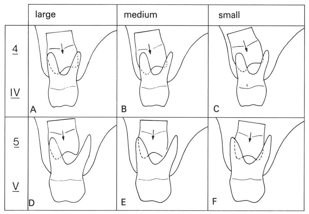
Fig. 6-4 Variations in the transverse position and direction of eruption of the maxillary first and second premolars related to the size of the middle section of the apical area. A shortage of space in the middle section of the apical area will affect the position and direction of eruption of the first premolar more than that of the second premolar.
A In a large middle section of the apical area the first premolar will be positioned superior to the first deciduous molar and its path of eruption will be slightly palatally directed.
B In a medium middle section of the apical area the first premolar will be more buccally placed and erupt in a more palatal direction.
C In a small middle section of the apical area the crowded conditions lead to excessive buccal displacement and a palatal direction of eruption.
D In a large middle section of the apical area the second premolar is located superior to its predecessor and more buccally. It will erupt in a slightly palatal direction.
E In a medium middle section of the apical area the second premolar is situated more central and superior to its predecessor. Its direction of eruption is about perpendicular to the occlusal plane.
F In a small middle section of the apical area the second premolar is more palatally located and erupts in a slightly buccal direction.
In general there will be a shortage of space in the middle section of the apical area in the canine-first premolar region. The second premolar is usually less affected because its predecessor has a larger mesiodistal crown dimension. However, an exception has to be made for cases of a mesially angulated erupting first permanent molar in which the distal surface of the second deciduous molar can resorb, leading eventually to a premature loss of that tooth (see Chapter 16). The transition of the posterior teeth that takes place some years later will then encounter special difficulties. This is also the case for premature loss of the second deciduous molar due to caries or extraction. The same holds true for the first deciduous molar, though to a lesser extent (see Chapter 14).
In a small middle section of the apical area the second premolar will be more distally located in relation to its predecessor without any deviation in its mesiodistal orientation. The mesially angulated first premolar and canine will erupt more mesially than normal.
Frequently, the space available between the first premolar and the root of the lateral incisor for eruption of the canine is limited. The first premolar will move distally when space in the dental arch and occlusion allow this migration to occur. Further, the position of the lateral incisor will change because the large canine crown will descend in close proximity to its root, which increases in circumference from the apex to the cementoenamel junction. Before emergence of the canine, the root of the lateral permanent incisor will be displaced mesially and palatally and its crown tipped distally and laterally. The extent of this movement of the lateral incisor is related to the lack of space encountered by the erupting canine and the presence or absence of the deciduous canine.
In exceptional instances the crown of the canine will be located not labially but palatally to the root of the lateral incisor. When adequate space is available the eruption path of the canine will />
Stay updated, free dental videos. Join our Telegram channel
VIDEdental - Online dental courses


