SECTION 6
TOOTH RESTORATION WITH CROWNS, BRIDGES OR VENEERS
REASON FOR PROCEDURE
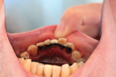
Each time a tooth is restored with a filling some of the tooth tissue is removed. Eventually, this will compromise the strength of the remaining tooth and it may begin to fracture under normal occlusal forces. This occurs especially when teeth have been root treated, and so it is usual for heavily filled and root-filled teeth to be crowned before fracture occurs.
In other cases, a tooth may be poorly shaped and require elective crowning to be more aesthetically pleasing. Similarly, a tooth may be too poorly shaped to assist in the retention of, for example, a denture, but this can be made possible by elective crowning.
CROWNS
Background information of procedure
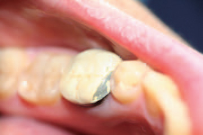
Posterior crowns are usually constructed from non-precious or precious metals, such as gold (see Figure 6.1). These provide maximum strength to withstand occlusal forces and have no risk of fracture. Anterior crowns are made of either tooth-coloured ceramic or porcelain throughout, or have porcelain bonded to a substructure of metal, and these give an aesthetically pleasing result when shaded and matched accurately to the adjacent teeth. Although modern techniques of crown construction are superb, it is possible for ceramic crowns to fracture or break away from their metallic substructure, so that repairs or even replacements are required. This can occur in patients with especially heavy bites or in those who grind their teeth (see Figure 6.2).
Figure 6.1 Full gold crown
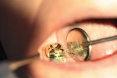
Figure 6.2 Fractured porcelain bonded crown
Details of procedure
Unless the tooth to be crowned has been root treated and is therefore nonvital, crown preparation is usually carried out under local anaesthetic so that the patient feels neither pain nor thermal stimulation in the tooth throughout the procedure.
As the crown must be individually constructed by a technician, the prepared tooth will require thermal protection in the meantime by being covered with a temporary crown material, such as acrylic.
During the preparation procedure, a dental nurse maintains good moisture control in the oral cavity using high-speed suction equipment. This provides a clear field of vision for the dentist, as well as making the patient more comfortable by removing water, saliva and tooth debris from the mouth.
Technique
- The dentist, nurse and patient wear personal protective equipment
- Local anaesthetic is administered and allowed to take full effect
- The shade and shape of crown are chosen by the dentist and patient
- A rubber dam is placed if required
- All sides and occlusal surfaces of the tooth are reduced by a uniform amount using a burr in the high-speed handpiece, so that space is created for the crown to be constructed and fitted without altering the patient’s occlusion
- The side reduction is completed to produce a near-parallel tooth core in order to give maximum retention of the crown on cementation
- Once tooth preparation is complete, impressions are taken of both arches and the patient’s normal biting position is recorded
- The impression of the opposing arch can be taken in alginate, but that of the working arch must be in a very accurate, non-tearing, elastomeric material, such as a silicone or polyether
- When satisfactory impressions have been produced, the tooth core is coated with a temporary acrylic material to prevent sensitivity and to restore some degree of aesthetics whilst the permanent crown is constructed
- Once the crown has been constructed, the patient reattends for its cementation
- Again, local anaesthetic is administered and a rubber dam is placed if required
- On removal of the temporary material, the crown is placed onto the tooth core and checked for accuracy of fit, shade and occlusion
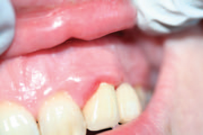
- If satisfactory, the crown is cemented permanently onto the tooth core using one of a variety of luting cements, taking great care to ensure all excess cement is removed (see Figure 6.3)
Figure 6.3 Localised gingivitis owing to incomplete cement removal at crown fit
AFTERCARE OF CROWNS
As with fillings, microscopically, all fixed prosthetic restorations provide a surface area for the attachment of plaque and oral bacteria. As crown margins are deliberately placed at the gingival margin to give superior aesthetics, any plaque accumulation can potentially cause either caries of the underlying tooth core or periodontal disease down the root of the tooth (see
Stay updated, free dental videos. Join our Telegram channel
VIDEdental - Online dental courses


