6: Genetics

Genetics

Heddie O. Sedano

Objectives

After studying this chapter, the student will be able to:

1. Define each of the words listed in the vocabulary for this chapter.

2. State the purpose of mitosis.

3. State the purpose of meiosis.

4. Explain what is meant by the Lyon hypothesis and give an example of its clinical significance.

5. Explain what is meant by a gross chromosomal abnormality and give three examples of syndromes that result from gross chromosomal abnormalities.

6. List the four inheritance patterns described in this chapter.

7. Explain what is meant by X-linked inheritance.

8. State the inheritance pattern and describe the oral manifestations and, if appropriate, the characteristic facies for each of the following: cyclic neutropenia, Papillon-Lefèvre syndrome, cherubism, Ellis–van Creveld syndrome (chondroectodermal dysplasia), mandibulofacial dysostosis (Treacher Collins syndrome), osteogenesis imperfecta, hereditary hemorrhagic telangiectasia (Osler-Rendu–Parkes Weber syndrome), Peutz-Jeghers syndrome, white sponge nevus (Cannon disease), hypohidrotic ectodermal dysplasia, hypophosphatasia, and hypophosphatemic vitamin D–resistant rickets.

9. State the inheritance pattern, the oral or facial manifestations, and the type and location of the malignancy associated with each of the following syndromes: Gardner syndrome; nevoid basal cell carcinoma syndrome (Gorlin syndrome); multiple mucosal neuromas, medullary carcinoma of the thyroid gland, and pheochromocytoma syndrome (multiple endocrine neoplasia type 2B [MEN 2B]); and neurofibromatosis of von Recklinghausen.

10. State the location and malignant potential of the intestinal polyps in Peutz-Jeghers syndrome and Gardner syndrome.

11. List the four types of amelogenesis imperfecta.

12. Briefly compare and contrast dentinogenesis imperfecta, amelogenesis imperfecta, and dentin dysplasia, including the inheritance patterns, the clinical manifestations, and the radiographic appearance of each.

Vocabulary

Genetics is the science that studies inheritance and the expression of inherited traits. The main objectives of this chapter are to introduce some of the basic concepts of genetics and present the clinical manifestations of some inherited oral disorders of interest to the dental hygienist. The descriptions of many syndromes are included. As explained in previous chapters, a syndrome is a distinctive association of signs and symptoms occurring together in the same patient. The syndromes included in this chapter are inherited. However, other syndromes such as acquired immunodeficiency syndrome (AIDS) are acquired, not inherited. In addition, the fact that an alteration is found as part of a syndrome does not mean that it cannot also occur independently. For example, cleft lip and palate occur as components of several syndromes and can also occur independently. The classification of syndromes is difficult because they are composed of several associated anomalies and the anomalies that compose the syndrome may not be present consistently in all patients with the same syndrome.

The term phenotype is used often in this chapter. It refers to the physical, biochemical, and physiologic traits of an individual. A phenotype can occur as a result of genetic factors or from a combination of genetic factors and environmental influences.

In this chapter, as in previous chapters, basic concepts are discussed first. These concepts are followed by descriptions of specific inherited disorders.

Chromosomes

The hereditary units that are transmitted from one generation to another are called genes. They are found on chromosomes, which are located in the nucleus of the cell. Using a microscope, one can see chromosomes clearly only when the nucleus and cell are dividing (Figure 6-1). At other times the genetic material is dispersed in the nucleus (see Figure 6-1). Each cell of the human body, with the exception of mature germ cells (ova and spermatozoa), has 46 chromosomes. Half of these chromosomes are derived from the father, and the other half from the mother.

Chromosomes contain deoxyribonucleic acid (DNA), which directs the production of amino acids, polypeptides, and proteins by the cell. In addition, DNA has the ability to duplicate itself (self-replication). It creates exact copies of itself; and through the process of cell division cells identical to the original cell are formed.

Normal Cell Division

Mitosis

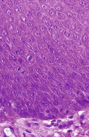
All cells in the body, with the exception of ova and spermatozoa, are called somatic cells. Cellular division is achieved by mitosis during a part of the life span of the somatic cell, called the mitotic cycle. The function of mitosis is to create an exact copy of each chromosome and, through division of the original cell, distribute an identical set of chromosomes to each daughter cell. After each cell division is completed and before the next division can occur, the cell enters the gap 1 (G1) phase, which is followed by the S phase, in which replication of the DNA takes place. The gap 2 (G2) phase follows the S phase and ends when mitotic division begins. The cell cycle is illustrated in Figure 6-2.

Stages of Mitosis

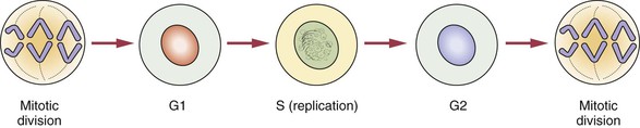
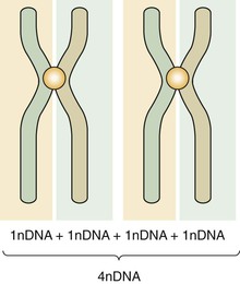
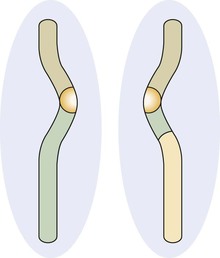
Mitosis is composed of four stages: (1) prophase, (2) metaphase, (3) anaphase, and (4) telophase. In each of these four stages, the chromosomes are distributed in a specific arrangement. In metaphase the chromosomes stain intensely and are arranged almost symmetrically at both sides of the center, or equatorial plane, of the cell. The appearance of a metaphase chromosome resembles the letter X (Figure 6-3), having a pair of “long arms” also known as q arms (see Figure 6-3, 3) and a pair of “short arms” also known as p arms (see Figure 6-3, 1). The size of the chromosome at metaphase and the length of the long and short arms vary from chromosome to chromosome. The constriction present in all chromosomes, which joins the short and long arms, is called the centromere (see Figure 6-3, 2). During metaphase chromosomes are actually formed by two identical vertical halves, each composed of either left or right short and long arms and half of the centromere. Each of these identical halves is called a chromatid (see Figure 6-3, 4). At metaphase each chromatid contains one molecule of DNA; therefore the DNA content of each chromosome is doubled (Figure 6-4). When cell division takes place, each chromosome splits vertically at the centromere; 46 chromatids (which now become chromosomes) form one daughter cell, and the other 46 chromatids form a second daughter cell. During prophase the chromosomes are lining up toward metaphase; in anaphase and telophase the chromatids are in the process of splitting.

Meiosis

Primitive germ cells (oogonia, spermatogonia) have 46 chromosomes. Mature germ cells (ova, spermatozoa) have 23 chromosomes. Meiosis is a two-step special type of cell division in which the primitive germ cells reduce their chromosome number by half and become mature germ cells. The primitive germ cells have two chromosomes for each pair and are called diploid. The suffix -ploid refers to the number of sets of chromosomes, and its prefix refers to the degree of ploidy. In diploid di- indicates two. The mature germ cells (or gametes) have half the number of chromosomes and are called haploid. During the period in which the cell is not in division, the DNA content of diploid cells is designated 2n DNA; in metaphase it is double or 4n DNA. After the two stages of meiosis have been completed, the 4n DNA is reduced to 1n DNA. The two steps are called first meiosis and second meiosis. This reduction is necessary to maintain the normal number of human chromosomes. A new embryo must have 46 chromosomes per cell, as did its parents. Therefore the union of germ cells needs to result in 46 chromosomes. If two cells with 46 chromosomes were combined, the resulting cell would have 92 chromosomes.

First Meiosis

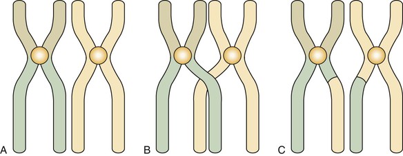
Before the first meiosis in the primitive germ cells, a replication of DNA occurs that is similar to that observed in the S phase of somatic cells. After replication the members of each pair of chromosomes line up next to each other in an intimate point-by-point relationship (Figure 6-5, A). This pairing does not occur in mitosis. After pairing, the two chromosomes establish actual contact at different locations. These contacts are known as chiasmata (meaning X shaped) and determine points of crossing over (Figure 6-5, B). This crossing over achieves the exchange of chromosome segments between a chromatid of one chromosome and a chromatid of the other chromosome of a pair (between homologous chromosomes) (Figure 6-5, C). This special aspect of the first meiosis takes place at metaphase. After metaphase of the first meiotic division, the chromosomes separate from each other, but no splitting of the centromere occurs. The chromosomes remain intact, and each member of the pair migrates to one of the new cells, each of which contains 23 chromosomes but twice the final amount of DNA. During this migration chromosomes of the paternal and maternal lines segregate at random, thus ensuring diversity of the species by creating a new combination of chromosomes.

On occasion, the chromosomes that were crossing over do not separate, and both migrate to the same cell. This is known as nondisjunction and results in the formation of a germ cell with an extra chromosome. If this occurs and that cell (either an ovum or a spermatozoon) participates in the formation of an embryo, three chromosomes (trisomy) instead of two result. An example of this type of abnormality is Down syndrome, also called trisomy 21, in which three of chromosome 21 are found instead of two. Trisomy has been reported for several different chromosomes.

In a female embryo oogenesis (ovum development) starts around the third month of prenatal life, and the future ova remain suspended, crossing over from about the time of birth until the time ovulation starts. At the beginning of ovulation the first meiosis is completed. Nondisjunction is more prevalent in female oogenesis than in male spermatogenesis. This is probably because of the period of prolonged crossing over; therefore the older the woman, the greater the chance of shedding a trisomic ovum and of bearing a child with Down syndrome or other trisomy.

Second Meiosis

The second stage of meiosis is essentially a mitotic division in which each chromosome splits longitudinally. No replication of DNA occurs before the second meiosis. After the splitting, two cells are formed, each containing the right amount of DNA (1n DNA) (Figure 6-6).

Nondisjunction can occur during the first and second meiosis. If it occurs in second meiosis, a chromosome does not split, and one daughter cell has a full chromosome, and the other has none.

Immediately after fertilization the chromosomes of the ovum and spermatozoon, each having 1n DNA, condense independently and form round structures, each known as a pronucleus. The DNA in these pronuclei replicates, forming a set of 23 full chromosomes for each, the maternal and paternal pronuclei. When the membranes of these pronuclei break, the 46 chromosomes mix at random, initiating the first cellular division (mitosis) that starts the development of the new embryo.

Lyon Hypothesis

The sex chromosomes are designated XX in women and XY in men. During the early period of embryonic development (possibly by the end of the second week), the genetic activity of one of the X chromosomes in each cell of a female embryo is inactivated. The inactivation is a random process affecting either the X chromosome derived from the mother or the X chromosome derived from the father. Activated chromosomes are dispersed in the nucleus. The inactivated chromosome remains contracted when the cell is not dividing and forms a structure known as the Barr body.

Barr bodies are only seen in female cells. The Barr body can be seen easily under the light microscope, especially in cytologic smears, including those obtained from the oral mucosa. The Barr body appears as a dark dot at the periphery of the nucleus (Figure 6-7).

This inactivation of one of the X chromosomes in a female embryo was postulated by Mary Lyon and is known as the Lyon hypothesis. This hypothesis has interesting clinical implications for female carriers of conditions caused by genes located on the X chromosome, which are explained later in this chapter.

Molecular Composition of Chromosomes

Deoxyribonucleic Acid

Chromosomes contain deoxyribonucleic acid (DNA). DNA contains the basic code or template that carries all genetic information. The basic unit of DNA is called a nucleotide, which is formed by a nitrogen-containing base, a five-carbon sugar (deoxyribose), and a phosphate. Four bases are found in DNA: adenine (A), guanine (G), thymine (T), and cytosine (C). These chains of polynucleotides are coiled to form a structure called a double helix (Figure 6-8). In DNA the base adenine is always bound to the base thymine, and guanine is always bound to cytosine. This is a consistent arrangement and is identical in all species from bacteria to humans, with just a few exceptions. Therefore the ratio of adenine to thymine (A/T) is always equal, and the same is true for the ratio of guanine to cytosine (G/C). In humans G/C pairs are about four times more frequent than A/T pairs. In Figure 6-8, the polynucleotide chains run vertically in opposite directions. Therefore a sequence of adenine, guanine, and cytosine (AGC) is always matched by the opposing sequence of thymine, cytosine, and guanine (TCG).

In Figure 6-8, the horizontal steps of the polynucleotide chains are nucleotides. Each pair of nucleotides is joined by a hydrogen bond, indicated by the dotted lines This arrangement is repeated horizontally to form the polynucleotide double spiral staircase (or helix) appearance of DNA (see Figure 6-8).

Each sequence of three bases is called a codon. It encodes an amino acid. Several amino acids form a polypeptide, and one or more polypeptides form a protein. A gene is often equated with the unit that forms a polypeptide.

DNA has the unique capability of self-replication, which is achieved by unwinding its double chain like the plaits of a braid. Each separated chain serves as a blueprint for another chain.

Mitochondrial DNA is found in the circular chromosome of the mitochondria, and it is maternally inherited. This is the DNA present in the cytoplasmic mitochondrial organelles of the ovum. Mitochondrial DNA is passed from the mother to all her offspring, regardless of sex.

Ribonucleic Acid

To produce amino acids, polypeptides, and proteins, the genetic code contained in the DNA is transcribed into ribonucleic acid (RNA), which differs from DNA in that it is a single strand (in its simplest form), its sugar is a ribose (the sugar in DNA is deoxyribose), and the base uracil (U) replaces the thymine (T) in DNA.

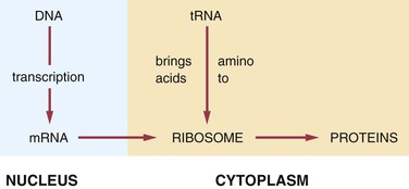
Types of Ribonucleic Acid

The four types of RNA are (1) messenger RNA (mRNA), (2) transfer RNA (tRNA), (3) ribosomal RNA (rRNA), and (4) heterogeneous nuclear RNA (hnRNA). RNA can be found in both the nucleus and the cytoplasm of a cell.

The first type of RNA, mRNA, is a blueprint of the genetic DNA for the coding of proteins. It carries the message for the DNA to ribosomes in the cytoplasm, in which proteins are produced. The second type of RNA, tRNA, transfers amino acids from the cytoplasm to the mRNA, positioning amino acids in the proper sequence to form polypeptides and hence proteins. The third type of RNA, rRNA, combines with several polypeptides to form ribosomes. The fourth type of RNA, hnRNA, is found within the nucleus and is the precursor of mRNA.

In the production of a protein (Figure 6-9) the mRNA carries the genetic code for the formation of that protein to the ribosomes. The tRNA brings amino acids to the ribosomes from the cellular cytoplasm. The amino acid sequence forms proteins according to the genetic code, and these proteins exit the ribosomes as they are formed.

Genes and Chromosomes

Genes in a chromosome are located in a linear manner. The genes in both members of a pair of chromosomes (homologous chromosomes) govern the same functions or dictate the same characteristics. The genes that are located at the same level (or locus) in homologous chromosomes and that dictate the same functions or characteristics are called alleles.

The manifestations (phenotype) of a gene action are not necessarily the same from one individual to another. This is best explained with the ABO blood group system. The locus can be occupied by either the factor that determines the blood group A or the factor that determines the blood group B. If it is empty, it results in the blood group O. The locus is always the same, and the three genes govern the same function; however, the clinical result is different. In this situation the trait or condition is said to have multiple alleles. For example, if both loci are AA or if they are AO, the person is said to have blood group A. If both loci are BB or BO, the person is said to have blood group B. If the loci are AB or BA, the person is said to have blood group AB. Only if both the loci are empty does the person have blood group O. The locus always controls the blood group, but the group depends on the alleles that are present or lacking in each person.

When the allelic genes are identical, the person is said to be homozygous for that gene, or a homozygote. Using the ABO blood group system again as an example, a person with AA, BB, or OO would be homozygous. When the genes are different (e.g., AB, AO, or BO), the person is said to be heterozygous for that gene, or a heterozygote. If a gene can express its effect clinically with a single dose (heterozygous), as in the combination AO = blood group A, the characteristic is said to be dominant. If the gene needs a double dose to exhibit its action (homozygous), the resulting characteristic or function is said to be recessive. For example, only the combination OO results in the blood group O.

Chromosomal Abnormalities

Abnormalities of chromosomes can be divided into two categories: (1) molecular abnormalities and (2) gross abnormalities. Molecular alterations occur at the DNA level and are not detectable microscopically. Most inherited disorders represent examples of molecular changes (mutations) at the level of one or both allelic genes. Examples of these conditions are presented later in this chapter.

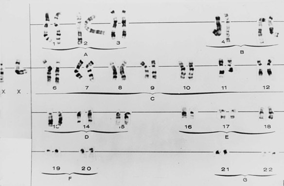
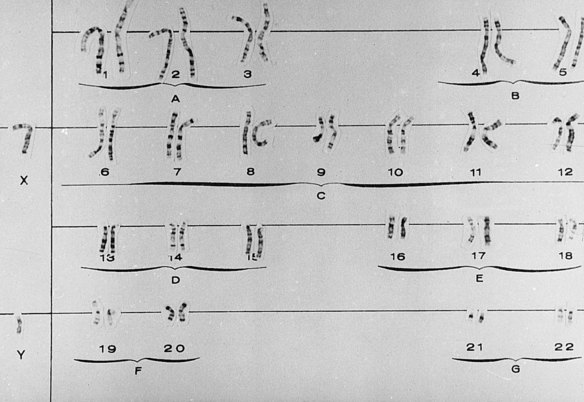
Gross chromosomal alterations can be observed in a karyotype. A karyotype (Figures 6-10 and 6-11) is a photographic representation of a person’s chromosomal constitution. Clinicians can create a karyotype by culturing cells from blood, skin, or other tissues. One method uses peripheral blood by placing it in a test tube with heparin to avoid coagulation and centrifuging it. After centrifugation the white blood cells (leukocytes) are deposited at the bottom of the tube. The leukocytes are removed and placed in a culture medium containing phytohemagglutinin, which is a substance that enhances mitosis. Because chromosomes are best observed when cell division is arrested at metaphase, colchicine is added to the culture after 72 hours of culture at 37° C to stop mitosis at metaphase. This also prevents the centromere from dividing. A hypotonic solution is then added to the culture to make the cells swell. The cells are then fixed (preserved) and stained and observed under the microscope. Examples of well-defined mitoses are chosen and photographed. The photograph is enlarged, and each chromosome is cut out of the photographic print. When stained, the chromosomes have a bandlike appearance that allows an accurate identification of each. These cutouts are then pasted on a special chart to construct the karyotype.

Gross Chromosomal Abnormalities

Alterations in Number and Structure of Chromosomes

Gross chromosomal abnormalities are caused by either alterations in chromosome number, which are almost always a result of nondisjunction (explained earlier), or alterations in structure, which develop because of chromosomal breaks or abnormal rearrangements. The following illustrate alterations in number:

Examples of structural abnormalities include the following:

Clinical Syndromes Resulting From Gross Chromosomal Abnormalities
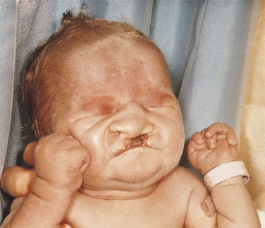
Trisomy 21

Trisomy 21, also known as Down syndrome, is the most frequent of the trisomies. Ninety-five percent of cases of Down syndrome are the result of nondisjunction, mostly associated with late maternal age at the time of conception.

Slanted eyes characterize the facies (the appearance of the face). Patients are generally shorter than normal, and heart abnormalities are present in more than 30% of individuals with trisomy 21. The intelligence level varies from near normal to markedly low.

Fissured tongue is frequently seen in patients with trisomy 21. Premature loss of teeth, especially the mandibular central incisors, caused by alveolar bone loss is seen frequently. Gingival and periodontal disease has been reported in 90% of affected individuals. Hypodontia (fewer teeth than normal), abnormally shaped teeth, and anomalies in eruption with malposition and crowding of teeth are common findings. Dental hygienists play an important role in the maintenance of oral health in these patients.

Trisomy 13

Trisomy 13 is characterized by multiple abnormalities in various organs. Seventy percent of live-born infants die within the first 7 months of life. Characteristic clinical findings include bilateral cleft lip and palate, microphthalmia (small eyes) or anophthalmia (no eyes), superficial hemangioma of the forehead or nape of the neck, growth retardation, severe mental handicap, polydactyly of hands and feet (supernumerary digits), clenching of the fist with the thumb under the fingers, rocker-bottom feet, heart malformations, and several anomalies of the external genitals. The facial appearance is quite striking because of the cleft lip, cleft palate, and ocular abnormalities (Figure 6-12).
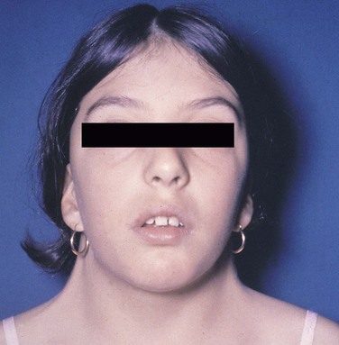
Turner Syndrome

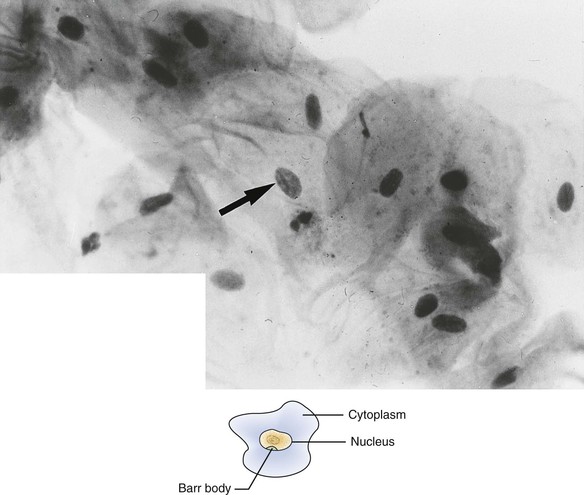
Patients with Turner syndrome have a female phenotype, and in the majority of cases the karyotype has the normal 44 autosomal chromosomes and only one X chromosome. A normal female would have two X chromosomes: one from the mother and one from the father. Most cases of Turner syndrome are the result of nondisjunction of the X chromosome in the paternal gamete. Clinically, these women are of short stature and have webbing of the neck and edema of the hands and feet (Figure 6-13). They frequently exhibit a low hairline on the nape of the neck. The chest is broad with wide-spaced nipples. The aorta frequently is abnormal, and body hair is sparse. The external genitals appear infantile, and generally the ovaries are not developed; therefore these individuals have primary amenorrhea (abnormal temporary or permanent cessation of the menstrual cycle). Smears taken from the oral mucosa demonstrate the lack of Barr bodies.

Klinefelter Syndrome

Klinefelter syndrome occurs when an ovum carrying two X chromosomes is fertilized by a spermatozoon with a Y chromosome; therefore the fertilized ovum will have two X chromosomes plus a Y chromosome. The majority of cases result from nondisjunction of the X chromosome, generally in the ova of older women. Affected individuals have a male phenotype, and the condition cannot be detected clinically until after puberty. These patients are taller than normal and have wide hips and female pubic hair distribution. About 50% have gynecomastia (development of female breasts), and intelligence levels are lower than normal in 10% of affected individuals. The penis appears normal, but the testes are smaller and harder than normal and lack seminiferous tubules.

The maxilla is slightly hypoplastic (underdeveloped). Buccal smears reveal the presence of one Barr body.

Variations of Klinefelter syndrome also occur; they are represented by karyotypes containing XXXY or XXXXY. The greater the number of X chromosomes, the more pronounced the clinical manifestations, and the lower the level of intelligence. The maxilla becomes increasingly hypoplastic with increasing number of X chromosomes. Buccal smears show one Barr body for each extra X chromosome.

Patterns of Inheritance

Because loci are present in both autosomal and X chromosomes and because of a double- and single-dose effect, four possible inheritance patterns exist. Dominant genes need only a single dose, and recessive genes need a double dose. The inheritance patterns are autosomal dominant, autosomal recessive, X-linked dominant, and X-linked recessive. Autosomal chromosomes include all chromosomes except those that determine sex (X and Y). The Y chromosome participates only in the differentiation of the masculine gonads.

Autosomal-Dominant Inheritance

A condition having autosomal-dominant inheritance is transmitted vertically from one generation to the next. Males and females are equally affected. When a person has a gene for the condition, the risk of having an affected offspring is 50% for each pregnancy. Genetic risk is always a mathematical estimate of probability governed by chance. Therefore none, less than half, half, more than half, or all of the offspring could be affected by a condition that is transmitted by autosomal-dominant inheritance.

An individual can carry a gene with a dominant effect without presenting any clinical manifestations. This is referred to as lack of penetrance. This situation can be explained partially by the presence of modifying genes in the same or other chromosomes. The clinical manifestations in autosomal-dominant disorders frequently vary among affected individuals. This is known as variable expressivity. Penetrance refers to the number of individuals affected, and expressivity pertains to the degree to which an individual is affected.

Autosomal-Recessive Inheritance

As stated previously, individuals exhibiting an autosomal-recessive trait must be homozygous for the gene. Clinically normal parents of affected children are heterozygous, and both are carriers of the trait. They are not generally recognized as carriers until after the birth of an affected child. If the enzymatic defect is known, carriers can be recognized before the birth of a child by assessing levels of the responsible enzyme in clinically normal members of a family with the trait. For parents who are carriers of the same recessive trait, the risk of having an affected child is 25%, the risk of having a homozygous normal child is 25%, and the chance of having a heterozygous carrier is 50% for each pregnancy. As in other inheritance patterns, risk is a mathematical estimate of the probability of an event occurring. If both parents are homozygous (have two of the genes of the trait) for a recessive trait, they would be expected to be affected, and all their children would be affected equally because they would also be homozygous (have two of the same genes) for the trait. In humans this type of situation is quite rare because individuals />

Stay updated, free dental videos. Join our Telegram channel

Jan 12, 2015 | Posted by in Oral and Maxillofacial Pathology | Comments Off on 6: Genetics

VIDEdental - Online dental courses

Get VIDEdental app for watching clinical videos