6
Diagnostic Adjuncts 2

Caries Detection
Dental caries is the most prevalent lesion in the oral cavity that is diagnosed and treated in a dental practice. Caries describes both the lesion and disease process, and its incidence in industrialised countries has decreased over the last three decades. Specifically, interproximal or smooth surface caries has shown a greater decline than occlusal lesions. Consequently, occlusal caries presents a greater challenge for early detection and treatment to prevent progression into larger cavities. Ideally, caries diagnostic tests should fulfil the following criteria:
- Sensitivity – ability to truly determine presence of caries;
- Specificity – ensure that sound tooth is registered as negative;
- Accuracy – number of positive and negative tests should sum to the total examined tooth surfaces.
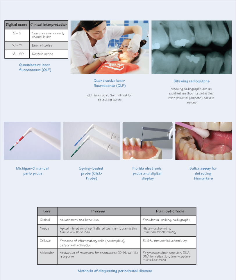
Methods for caries detection include the following.
- Histology – remains the gold standard for evaluating caries, but is obviously impractical in the dental surgery. Its use is limited to research, especially of the primary dentition following exfoliation or extraction.
- Visual inspection – most common method for caries diagnosis, e.g. using the International Caries Detection and Assessment System (ICDAS), but is affected by operator experience and subjectivity.
- Enhanced visual inspection includes:
Magnification – using loupes or operating microscope;Fibreoptic illumination – widely used for detecting interproximal caries, but can also be used for occlusal lesions, e.g. FOTI (fibreoptic transillumination) and DIFOTI (digital imaging fibreoptic transillumination). Sound enamel appears translucent, enamel caries appears grey and opaque, and dentine caries casts an orange/brown or bluish shadow;Enamel porosity (Ekstrand method) – assesses difference of refractive indices between air, water and enamel;Combination of fibreoptics and enamel porosity.
- Electrical conductance measurements (ECM) – measures electrical resistance of teeth. Intact enamel h/>
Stay updated, free dental videos. Join our Telegram channel

VIDEdental - Online dental courses


