53 Jaw bone conditions: Radiolucencies and radiopacities
Figure 53.1a Radiolucent lesions.
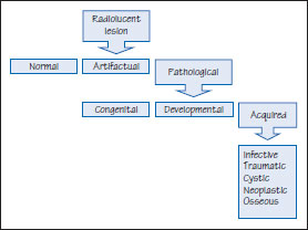
Figure 53.1b Nonodontogenic radiolucent cystic lesions.
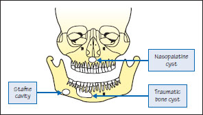
Figure 53.2 Nasopalatine duct cyst.
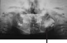
Figure 53.3 Hyperparathyroidism. Brown tumor.
Figure 53.4a Radiopaque lesions.
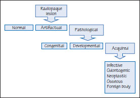
Figure 53.4b Radiolucent lesions. CEOT, calcifying epithelial odontogenic tumor; KCOT, keratocystic odontogenic tumor; AOT, adenomatoid odontogenic tumor.
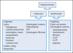
Figure 53.5 Osteosclerosis.
Figure 53.6a Osteoma.
Figure 53.6b CT Osteoma.
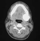
Figure 53.7 Osteosarcoma.
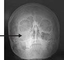
Figure 53.8 Ameloblastic fibro-odontoma.
Many diseases of or in the jaws present asymptomatically as radiolucencies, radiopacities or with mixed appearances on imaging. Other presentations are as swellings, pain or sometimes fracture or disturbance of tooth eruption (displaced, missing or loose teeth). Swellings that appear to originate from the jaws may arise from subcutaneous tissues or bones: the mnemonic MINT aids diagnosis:
Malformations, e.g. tori and fibro-osseous lesions
Inflammatory conditions, e.g. odontogenic infections, osteomyelitis, actinomycosis, tuberculosis, or syphilis
Neoplasms and cysts (see below)
Trauma causing subperiosteal hematomas
Investigations largely involve imaging, and serum calcium, phosphate, and alkaline phosphatase levels, but histopathology is almost invariably required (Tables 53.1 and 53.2).
Radiolucencies
Radiographic features to be assessed include the lesional size, shape, number, margins, character (radiolucency and/or radiopacity), and effects (displacement of the inferior alveolar nerve or tooth displacement or resorption).
Well-defined radiolucencies are often odontogenic cysts and benign tumors.
Poorly defined radiolucencies are often infections or tumors.
Jaw radiolucencies may include (Figures 53.1a and b):
- Odontogenic diseases, inflammation, cyst/>
Stay updated, free dental videos. Join our Telegram channel
VIDEdental - Online dental courses


