Chapter 5
The Future
Aim
To be appraised of emerging developments in the early diagnosis, monitoring, risk factor identification and prevention of dental erosion.
Outcome
After reading this chapter the practitioner should have an understanding of:
-
Emerging technologies of potential assistance in making an early diagnosis of dental erosion.
-
Potential future risk factor identification tools
-
New potential preventive regimes.
The Future
Once a diagnosis of dental erosion has been made practitioners face a number of choices. They may elect to institute a preventive regime, with monitoring of tooth surface loss, or to intervene operatively. A balance has to be struck between the risks of further tooth substance loss occurring, should prevention fail, and providing restorative treatment that inevitably will require maintenance and replacement. Should the wrong decision be made the patient will either be condemned to unnecessary restorative treatment or future restorative options will be denied them due to a lack of tooth structure. Fig 5-1 illustrates such a dilemma. Are there any developments that may assist the dentist and the patient, either emerging now or likely to do so in the future? Especially in relation to early diagnosis and monitoring, identification of risk factors and new preventive regimes.
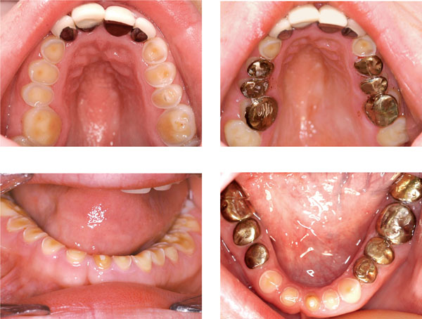
Fig 5-1 The dilemma of observing versus treatment – this extreme example illustrates how prolonged loss of tooth substance and zealous monitoring compromised tooth structure. Residual tooth structure was eventually protected by the provision of adhesively bonded gold onlays with no prior tooth preparation.
Early Diagnosis and Monitoring
Dental erosion arises from the loss of enamel and dentine from acid attack. It is therefore a consequence of the chemical process of demineralisation where gradual and sustained acid exposure progressively depletes the mineral content of the tooth. This ultimately culminates in a change in surface morphology. Much of this alteration in level of mineralisation occurs beneath the visible tooth surface. Eroded lesions are principally comprised of two distinct zones. The first is the defect formed by the actual loss of tooth substance. Beneath this is a layer of softened demineralised enamel/dentine that, given sufficient time and erosive challenge will be lost, contributing to further defect formation and a new softened tooth surface. It is relatively late in the erosive process when the tooth surface becomes changed in shape as surface softening must precede defect formation. Currently it is only then that the erosive process is highlighted to the dentist. If the early loss of mineral could in some way be detected before an alteration in surface morphology occurs preventive strategies could be put into place, that if successful, would conserve valuable tooth substance. No chairside technique currently exists to assess changes in mineral content, although in the laboratory quantitative light-induced fluoresence (QLF) has successfully been used to quantify mineral loss. In essence, sound coronal tooth substance, when subjected to a beam of monochromatic blue light of a wavelength of 370nm, fluoresces yellow-green. This can be measured and compared to eroded tissue where the scattering of the light, due to increased porosity, reduces the level of auto-fluoresence (Fig 5-2). Much development work however, still needs to be carried out on this useful laboratory tool if it is to develop into a chair-side test.
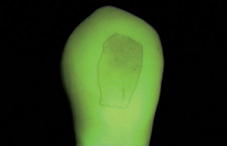
Fig 5-2 Laboratory generated erosion of human tooth as viewed using QLF. The area that is unaffected is the windowed area. This buccal area was protected from the erosive attack of orange juice by a coating of nail varnish.
Currently it is universally recognised that study models, recorded at sequential intervals, are useful for monitoring the progression of tooth surface loss and assessing the impact of preventive advice upon the erosive process. They are however, both bulky and fragile. Although valuable storage space may be saved in the surgery by issuing them to the patient for safe keeping they could well become lost or broken resulting in the loss of much valuable information. The use of digital study model capture and storage systems, although potentially rendering conventional study models obsolete in the future, is still in its infancy. Despite this the detection capacity of ongoing tooth surface loss, by comparing sequential study casts, is relatively gross and insufficiently sensitive to assess the subtle early impact of preventive measures – such as an alteration in dietary intake. This therefore misses an important opportunity to educate and motivate patients by illustrating to them that their change in lifestyle has slowed or ceased the rate of tooth surface loss. Within the research community, in recent years, there has been some use of surface mapping techniques to quantify the degree of tooth surface loss. Such measurement is made difficult by the lack o/>
Stay updated, free dental videos. Join our Telegram channel
VIDEdental - Online dental courses


