5: Orofacial Pain

Orofacial Pain

Pain Physiology

The International Association for Study of Pain (IASP) in 1994 defined pain as the subject’s conscious perception of modulated nociceptive impulses that generate an unpleasant sensory and emotional experience associated with actual damage or described in terms of such damage.

The experience of pain is usually a protective mechanism of the body. On a short-term basis, pain warns the individual that he or she is in danger so that one can alter the situation. For example when a person accidentally touches something hot he or she will alter the situation by spontaneously withdrawing from the source of injury. Long-term pain will result in immobilization of the affected part such that the individual can recover from the injury faster (for example, muscle spasm).

Pain at times can be non-beneficial, such as pain associated with cancer, psychogenic pains and neuralgias which only adds to the misery of the patient. However, interestingly these pains help the physician in diagnosis.

Properties of Pain

Weber and Fechner’s law

Weber and Fechner proposed that gradation of stimulus strength is discriminated approximately in proportion to the logarithm of stimulus strength.

This law can be mathematically expressed as

image

where R = intensity of the reaction (i.e. the pain perceived),

α = constant and

S = the intensity of the stimulus.

According to this law, exponential increase in the intensity of stimulus does not cause exponential increase in pain perceived. Though the pain perceived increases with increase in stimulus; the pain experienced is only in terms of log of the intensity of the stimulus.

For example, if a pin prick (1 unit of tissue damage) of 1 unit of intensity of stimulus results in 1 unit of pain perception; a musculocutaneous laceration (100 units of tissue damage) causing 100 units of intensity of stimulus results in 2 units of pain perception and not 100 units. Similarly, a crushing injury (1,000 units of tissue damage) causing 1,000 units of intensity of stimulus results in 3 units of pain perception and not 1,000 units.

This law ensures that an individual would perceive pain of all intensities but at the same time will not cause significant morbidity due to pain.

Nociceptors

Pain receptors are called nociceptors (from Latin, nocere–to hurt). All nociceptors are free nerve endings (however other cutaneous receptors when stimulated excessively can result in pain). Either present in skin or any other tissues. They are more concentrated in superficial layers of skin, periosteum, arterial walls, joint surfaces, the falx and the tentorium of the cranial vault. Deep tissues are sparsely supplied with nociceptors.

Two general types of nociceptors are characterized by the neurons associated with them.

Aδ fiber C fiber
1–6 μm diameter 1.5 μm diameter
Myelinated Non-myelinated
5–30 m/s conduction velocity 0.5–2 m/s conduction velocity
Carries the first pain that is experienced–sharp Carries steady dull pain

Stimulation of nociceptors

Mechanical, chemical and thermal stimuli excite pain receptors. Fast pain is conducted by Aδ fibers which is elicited by the mechanical and thermal stimuli. The slow dull pain is conducted by the C fibers which are elicited by all three types of stimuli. It is almost always caused by release of chemicals liberated by the injured tissue. These are endogenous chemicals called algogenic (pain producing) substances. Algogenic substances stimulate nociceptors to produce pain.

These chemicals are pain producing peptides, bradykinins, serotonin (5 HT), potassium ions, prostaglandins, acetylcholine and proteolytic enzymes.

It is interesting to note that commonly used NSAIDs suppresses pain by inhibiting prostaglandin synthesis. These prostaglandins by themselves cannot excite the nociceptors, i.e. prostaglandins are not algogenic but they enhance the sensitivity of the nociceptors toward the algogenic power of bradykinins and other chemical mediators of pain.

Sequelae of Pain

Apart from nociception there are various other sequelae of pain that alter other systems of the body. Pain affects an individual psychologically as well as physically.

Dual Pain Pathways

Peripheral pain fibers are of two types, the fast conducting (Aδ fibers) and slow (C fibers) conducting. The fast pain is felt within 0.1 second of the application of the noxious stimulus. But it takes 1 second or more for slow pain to begin. Occasionally slow pain may take over a few minutes to begin after application of the noxious stimulus.

Though both fast and slow conducting fibers have free nerve endings as their receptors, they travel via two distinct pathways for transmitting pain signals to the central nervous system. Because of this dual pain pathway a single painful stimulus would often give a first sharp electrical pain that is generally followed by a slow dull pain. This ‘double’ pain sensation initially would cause an individual to react immediately to safeguard himself/herself. Whereas the slow pain tends to become increasingly painful over a period of time.

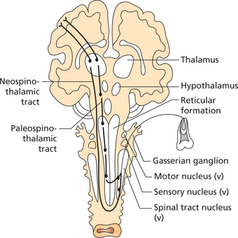
Once these fibers (Aδ and C) enter the spinal cord, the pain signals take two pathways to the brain. They are the neospinothalamic tract and the paleospinothalamic tract (Figure 1).

Neospinothalamic tract (for fast pain)

Sensations of mechanical and acute thermal pain are brought to the spinal cord by the first order neurons. They terminate in the lamina I (lamina marginalis) in the dorsal horns where they synapse to excite the second order neurons of the neospinothalamic tract. These fibers cross immediately to the opposite side through the anterior commissure and then travel upward in the anterolateral column of the spinal cord.

Few of these second order neurons terminate in the reticular areas of the brain stem (the reticular areas when stimulated causes excessive alertness and increases an individual’s sense perception), while most of the others travel up to the thalamus terminating in the ventrobasal complex along with the dorsal column–medial lemniscal tract. The remaining second order neurons terminate in the posterior nuclear group of the thalamus.

From these areas third order neurons relay signals to other basal areas of the brain and to the somatic sensory cortex.

Analgesia system or pain inhibitory pathway

The analgesia system comprises three major components, namely,

The electrical stimulation in the periaqueductal gray area or in the raphe magnus nucleus can almost completely suppress many strong pain signals entering the dorsal spinal roots. Stimulation of areas at still higher levels in the brain that in turn excite the periaqueductal gray can also suppress pain.

The nerve fibers originating from periaqueductal gray and periventricular nuclei secrete enkephalin at their terminals. These fibers synapse with the fibers originating from the raphe magnus nucleus.

The fibers originating from the raphe magnus nucleus secrete serotonin at their nerve terminals. Finally these fibers terminate at the dorsal horns of the spinal cord. Serotonin (secreted by the fibers originating from the raphe magnus nucleus) causes the cord neurons to secrete enkephalins.

Enkephalin causes presynaptic inhibition and post synaptic inhibition of Aδ and C pain fibers at the site of synapse of the dorsal horns. This inhibition is caused by the blocking of calcium channels of nerve membrane at their terminals.

Gate Control Theory

This theory explaining pain modulation was proposed by Ronald Melzack and Patrick David Wall in 1965.

They hypothesized that pain perception was not just solely due to activation of nociceptors but due to interaction between pain conducting and non-pain conducting neurons. Non-pain conducting nerve fibers interfere with the pain conduction thus altering pain perception.

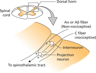
There have been various modifications of the original gate control theory. A revised version of the gate control hypothesis proposes that the projection neuron of the spinothalamic pathway when activated results in sensation of pain (Figure 2).

The projection neuron synapses with both non-nociceptive mechanoreceptors (Aα and Aβ fibers) and the nociceptive C fibers, hence activated by both. The interneuron is spontaneously active and this activation of interneuron causes inhibition of projection neuron; thus inhibiting pain perception.

In turn, the interneuron synapses with the projection neuron, C fibers, Aα and Aβ fibers. When the non-nociceptive mechanoreceptors (myelinated and fast conducting Aα and Aβ fibers) are stimulated they cause two effects: (i) activation of interneuron and (ii) activation of projection neuron.

Though it activates the projection neuron simultaneous activation of interneuron causes inhibition of projection neuron thus resulting in lack of pain sensation.

When C fibers are activated they again cause two effects: (i) inhibition of interneuron and (ii) activation of projection neuron, therefore resulting in pain perception.

When both non-nociceptive mechanoreceptors and nociceptors are simultaneously activated, the mechanoreceptors being myelinated fast conducting fibers, the signals from these fibers reach interneuron and projection neuron first before C fibers can conduct signals. In these instances the Aα and Aβ fibers stimulate the interneuron before C fibers can inhibit the same. Thus the net effect is no pain.

This would explain the benefits of physiotherapy and massage in the effective management of painful conditions.

It is interesting to note that free nerve endings of unmyelinated nerve fibers when relatively mildly stimulated produce itch and tickle.

Unlike pain, itching occurs in superficial tissues and not in deep structures like viscera.

It is common knowledge that scratching relieves itching as the fast conducting mechanoceptive fibers activated by scratching suppress the itching sensation.

Studies indicate that the C fiber system responsible for itching may not be the same responsible for pain. Surprisingly tickling sensation in general is regarded as pleasurable whereas itching and pain are regarded as unpleasant sensations.

Classification of Orofacial Pain

The American Academy of Orofacial Pain has classified orofacial pain as follows:

Bell (1989) has classified orofacial pain as follows:

Clinical Assessment of Pain

The diagnosis and management of orofacial pain begins with a comprehensive patient history. The most expeditious way to obtain such a history is via a detailed patient questionnaire and detailed patient-physician interview.

The interview is followed by a complete oral and head and neck examination, appropriate chair side investigations, radiographic imaging and lab studies.

Assessment of pain can be achieved by certain subjective and objective methods. Some widely accepted subjective methods include the use of McGill Pain Questionnaire (long/ short), visual analog pain scale (VAS), brief pain inventory. The faces pain scale and the pain diagram.

During the patient interview the clinician should obtain the following data: mode of onset, duration, location of the pain, quality or character of the pain, intensity of the pain, frequency of the painful episodes, aggravating and relieving factors if any, radiation or referral patterns, any other associated symptoms and history of any medical consultations/use of medications for the same.

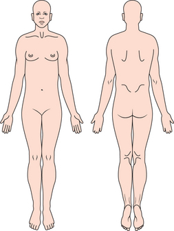
The location of pain and referral patterns can be identified by the patient using a pain diagram (Figure 3).
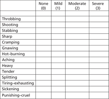
The pain quality or character can be expressed using the widely accepted McGill Pain Questionnaire (Figure 4). This questionnaire will help the patient to describe how exactly he/she feels about the pain by selecting an appropriate adjective from a list in the questionnaire.

The intensity of pain can be quantified using the visual analog scale (VAS). The VAS is a 10 cm long line with 0 marked on one end (represents no pain) and 10 at the other end (represents worst possible pain). The linear scale has markings from 0 to 10 at 1 cm intervals. The patient is encouraged to mark a point along this scale that correlates with the intensity of pain experienced. For convenience pain intensity can be categorized as mild (score 1–1), moderate (score 4–4) and severe for scores 7–7 (Figure 5).

However the ‘Faces Pain Scale’ can be used in child patients for assessing the intensity of pain (Figure 6).

Following the history, physical examination should be undertaken. A thorough physical examination of the head and neck, including the TMJ, maxillary sinus, masticatory muscles along with the accessory muscles, salivary glands and the oral cavity should be performed.

Intraoral examination should include the evaluation of teeth, periodo/>

Stay updated, free dental videos. Join our Telegram channel

Jan 12, 2015 | Posted by in Oral and Maxillofacial Radiology | Comments Off on 5: Orofacial Pain

VIDEdental - Online dental courses
Get VIDEdental app for watching clinical videos