5
Local anaesthesia for children
J.G. Meechan
Chapter contents
5.2.1 Intra-oral topical agents
5.2.2 Topical anaesthetics that will anaesthetize skin
5.2.3 Controlled-release devices
5.3 Non-pharmacological pain control
5.4 Local anaesthetic solutions
5.5 Techniques of local anaesthesia
5.5.1 Infiltration anaesthesia
5.5.2 Regional block anaesthesia
5.5.3 Intra-ligamentary anaesthesia
5.6 Pain-free local anaesthesia
5.7 Complications of local anaesthesia
5.7.1 Generalized complications
5.7.2 Early localized complications
5.7.3 Late localized complications
5.8 Contraindications to local anaesthesia
5.1 Introduction
This chapter considers the use of LA in children and describes methods of injection that should produce minimal discomfort. The complications and contraindications to the use of LA in children are also discussed. The major use of local anaesthetics is in providing operative pain control. However, it should not be forgotten that these drugs can be used as diagnostic tools and in the control of haemorrhage.
5.2 Surface anaesthesia
Surface anaesthesia can be achieved by physical or pharmacological methods (topical anaesthetics). One physical method employed in dentistry is the use of refrigeration. This employs the application of volatile liquids such as ethyl chloride. The latent heat of evaporation of this material reduces the temperature of the surface tissue and this produces anaesthesia. This method is rarely used in children as it is difficult to direct the stream of liquid accurately without contacting associated sensitive structures such as teeth. In addition, the general anaesthetic action of ethyl chloride should not be forgotten.
5.2.1 Intra-oral topical agents
The success of topical anaesthesia depends on the technique. Topical anaesthetic agents can anaesthetize a 2–3mm depth of surface tissue when used properly.
The following points should be noted when using intra-oral topical anaesthetics:
• the area of application should be dried;
• the anaesthetic should be applied over a limited area;
• the anaesthetic should be applied for sufficient time.
A number of different preparations varying in the active agent and in concentration are available for intra-oral use. In the UK the agents most commonly employed are lidocaine (lignocaine) and benzocaine. Topical anaesthetics are provided as sprays, solutions, creams, or ointments. Sprays are the least convenient as they are difficult to direct. Some sprays taste unpleasant and can lead to excess salivation if they inadvertently reach the tongue. In addition, unless a metered dose is delivered, the quantity of anaesthetic used is poorly controlled. It is important to limit the amount of topical anaesthetic used. The active agent is present in greater concentration in topical preparations than in local anaesthetic solutions and uptake from the mucosa is rapid. Systemic uptake is even quicker in damaged tissue. An effective method of application is to spread some cream on the end of a cotton bud (Fig. 5.1). All the conventional intra-oral topical anaesthetics are equally effective when used on reflected mucosa. The length of time of administration is crucial for the success of topical anaesthetics. Applications of around 15 seconds or so are useless. An application time of around 5 minutes is recommended. It is important that topical anaesthetics are given sufficient time to achieve their effect. This is especially important in children as this may be their initial experience of intra-oral pain-control techniques. If the first method encountered is unsuccessful, confidence in the operator and his armamentarium will not be established.
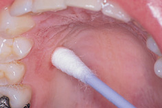
Figure 5.1 Use of a cotton bud to apply a topical anaesthetic over a limited area
Although the main use of topical anaesthetics is as a pre-injection treatment, these agents have been used as the sole means of anaesthesia for some intra-oral procedures in children including the extraction of mobile primary teeth.
5.2.2 Topical anaesthetics that will anaesthetize skin
EMLA® cream (a 5% Eutectic Mixture of the Local Anaesthetic agents prilocaine and lidocaine) was the first topical anaesthetic to be shown to produce effective surface anaesthesia of intact skin. Therefore it is a useful adjunct to the provision of general anaesthesia in children as it allows pain-free venepuncture.
When used on skin it has to be applied for about an hour so it is only appropriate for elective general anaesthetics. Clinical trials of the intra-oral use of EMLA® have shown it to be more effective than conventional local anaesthetics when applied to attached gingiva such as the hard palate and interdental papillae. It appears to be no more effective than conventional topical agents when applied to reflected mucosa. Intra-oral use of EMLA® is not recommended by its manufacturers. An intra-oral formulation of the combination of prilo-caine and lidocaine (Oraqix®) is available as a topical treatment for application to gingival pockets to help reduce the discomfort of dental scaling.
Tetracaine (amethocaine) 4% gel is another topical anaesthetic for skin that may be useful prior to venepuncture. Unlike EMLA,® which consists of amide local anaesthetics, tetracaine is an ester.
5.2.3 Controlled-release devices
The use of topically active agents incorporated into materials that adhere to mucosa and allow the slow release of the agent is a potential growth area in the field of local anaesthetic delivery. Such techniques might prove to be of value in paediatric dentistry. Clinical studies investigating the release of lidocaine from intra-oral patches have shown some promise. The use of anaesthetic agents incorporated into liposomes is also promising in this regard.
5.2.4 Jet injectors
Jet injectors belong in a category somewhere between topical anaesthesia and LA but will be discussed here for completeness. These devices allow anaesthesia of the surface to a depth of over 1cm without the use of a needle. They deliver a jet of solution through the tissue under high pressure (Fig. 5.2).
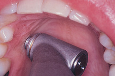
Figure 5.2 The jet injector. (Reproduced from Dental Update (ISSN 0305-5000), by permission of George Warman Publications (UK) Ltd.)
Conventional local anaesthetic solutions are used in specialized syringes and have been successful in children with bleeding diatheses where deep injection is contraindicated. Jet injection has been used both as the sole means of achieving LA and prior to conventional techniques. This method of anaesthesia has been used alone and in combination with sedation to allow the pain-free extraction of primary teeth. The use of jet injection is not widespread for a number of reasons. Expensive equipment is required, soft tissue damage can be produced if a careless technique is employed, and the specialized syringes can be frightening to children because of both their appearance and the sound produced during anaesthetic delivery. In addition, the unpleasant taste of the anaesthetic solution, which can accompany the use of this technique, can be off-putting.
Although no needle is employed, the technique is not painless. Indeed, careful use of conventional techniques has been shown to produce similar levels of discomfort to jet injection.
5.3 Non-pharmacological pain control
A number of non-pharmacological methods for reducing the pain of operative dentistry are now available, including the use of electrical stimulation and radio waves. Hypnosis also belongs in this category. There are reports that some lasers have the potential to produce LA as dental hard tissue can be removed painlessly by such devices. The use of refrigeration techniques was mentioned above.
Electroanalgesia or transcutaneous electrical nerve stimulation (TENS) has been shown to be effective in providing anaesthesia for restorative procedures in children aged 3–12 years. The technique has also been used to provide pain control during the extraction of primary teeth and as a ‘deep topical agent’ to reduce the pain of local anaesthetic injections. In younger children the level of stimulation is controlled by the operator. Children over 10 years of age can understand the method sufficiently well to be able to control the level of stimulus themselves.
TENS blocks transmission of the acute pain of dental operative procedures because large myelinated nerve fibres (such as those responding to touch) have a lower threshold for electrical stimulation than smaller unmyelinated pain fibres. Stimulation of these fibres by the current from the TENS machine closes the ‘gate’ to central transmission of the signal from the pain fibres. This is quite different from the use of TENS in the treatment of chronic pain, where the release of endogenous painkillers such as β-endorphins is stimulated. In addition, if the patient operates the machine, the feeling of control may help allay anxiety and aid in pain management.
Non-pharmacological methods of pain control offer two advantages. First, systemic toxicity will not occur and, second, the soft tissue anaesthesia resolves at the end of the procedure. This reduces the chances of self-inflicted trauma.
Hypnosis can be used as an adjunct to LA in children by decreasing the pulse rate and the incidence of crying. It appears to be most effective in young children.
5.4 Local anaesthetic solutions
A number of local anaesthetic solutions that can provide anaesthesia lasting from 10 minutes to over 6 hours are now available. There are few, if any, indications for the use of the so-called long-acting agents in children. The gold standard for many years has been lidocaine with adrenaline (epinephrine). Unless there is a true allergy to lidocaine, 2% lidocaine with 1:80 000 adrenaline is the solution of choice in the UK for most techniques. One technique where lidocaine with adrenaline is not the best choice is infiltration anaesthesia in the mandible. A number of studies in adults have shown that 4% articaine with adrenaline is superior for this method in both the molar and incisor regions. ‘Short-acting’ agents such as plain lidocaine are seldom employed as the sole agent because, although pulpal anaesthesia may be short-lived, soft tissue effects can still last for over an hour or so. More importantly, the efficacy of plain solutions is much less than those containing a vasoconstrictor.
5.5 Techniques of local anaesthesia
There are no techniques of local anaesthetic administration that are unique to children; however, modifications to standard methods are sometimes required. As far as positioning the child is concerned, the upper body should be around 30° to the vertical. Sitting upright can increase the chances of fainting, whilst at the other extreme (fully supine) the child may feel ill at ease. When there is a choice of sites at which to administer the first local anaesthetic injection, the primary maxillary molar area should be chosen. This is the region that is most easily anaesthetized with the least discomfort.
5.5.1 Infiltration anaesthesia
Infiltration anaesthesia is the method of choice in the maxilla. The infiltration of 0.5–1.0mL of local anaesthetic is sufficient for pulpal anaesthesia of most teeth in children. The objective is to deposit local anaesthetic solution as close as possible to the apex of the tooth of interest; however, the presence of bone prevents direct apposition. As the apices of most teeth are closer to the buccal side, a buccal approach is employed and the needle is directed towards the apex after insertion through reflected mucosa. Direct deposition under the periosteum can be painful; therefore a compromise is made and the solution is delivered supra-periosteally. The one area where pulpal anaesthesia can prove troublesome in the child’s maxilla is the upper first permanent molar region where the proximity of the zygomatic buttress can inhibit the spread of solution to the apical area (see below).
The use of buccal infiltration anaesthesia in the mandible will often produce pulpal anaesthesia of the primary teeth. However, it may be unreliable when operating on the permanent dentition, with the exception of the lower incisor teeth (Jaber et al. 2010). An alternative form of anaesthesia in the posterior mandible is inferior alveolar nerve block anaesthesia.
5.5.2 Regional block anaesthesia
Inferior alveolar and lingual nerve blocks
Administration of the inferior alveolar and lingual nerve block is easier to perform successfully in children than in adults. A common fault in adults is placing the needle too low on the ramus of the mandible with deposition of solution inferior to the mandibular foramen. In children, the mandibular foramen is low relative to the occlusal plane (Fig. 5.3), and it is difficult to place the needle inferior to this site if it is introduced parallel to the occlusal plane. Thus in children it is easier to ensure that the solution is deposited around the nerve before it enters the mandibular canal.
The technique of administration is identical to that used in adults and is best performed with the child’s mouth fully open. The direct approach (introducing the needle from the primary molars of the opposite side) is recommended as less needle movement is required after tissue penetration with this method compared with the indirect technique. The operator’s non-dominant hand supports the mandible with the thumb intra-orally in the retromolar region of the mandible. The index or middle finger is placed extra-orally at the posterior border of the ramus at the same height as the thumb. The needle is advanced from the primary molar region of the opposite side with the syringe held parallel to the mandibular occlusal plane. The needle is inserted through mucosa in the mandibular retromolar region lateral to the pterygomandibular raphe midway between the raphe and the anterior border of the ascending ramus of the mandible, aiming for a point halfway between the operator’s thumb and index finger. The height of insertion is about 5mm above the mandibular occlusal plane, although in young children entry at the height of the occlusal plane should also be successful. The needle should be advanced until the medial border of the mandible is reached. In young children bone will be reached after about 15mm and thus a 25mm needle can be used; however, in older children a long (35mm) needle should be employed as penetration up to 25mm may be required. Once bone has been touched, the needle is withdrawn slightly until it is supra-periosteal, aspiration is performed, and 1.5mL of solution is deposited. The lingual nerve is blocked by withdrawing the needle halfway, aspirating again, and depositing most of the remaining solution at this point. The final contents of the cartridge are expelled as the needle is withdrawn through the tissues. A common fault is to contact bone only a few millimetres following insertion. In most children this will lead to unsuccessful anaesthesia. This usually occurs because the angle of entry is too obtuse. If this happens, the needle should not be completely withdrawn but pulled back a couple of millimetres and then advanced parallel to the ramus for about 1cm with the barrel of the syringe over the mandibular teeth of the same side. The body of the syringe is then repositioned across the primary molars or premolars on the opposite side and advanced towards the medial border of the ramus.
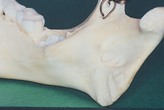
Figure 5.3 The mandibular foramen is below the occlusal plane in children. (Reproduced from Dental Update (ISSN 0305-5000), by permission of George Warman Publications (UK) Ltd.)
Long buccal and mental and incisive nerve blocks
The long buccal injection usually equates to a buccal infiltration in children. The mental and incisive nerve block is readily administered in children as the orientation of the mental foramen is such that it faces forward rather than posteriorly as in adults (Fig. 5.4). Thus it is easier for solution to diffuse through the foramen when approached from an anterior direction. The needle is advanced in the buccal sulcus and directed towards the region between the first and second primary molar apices. Blockade of transmission in the mental nerve provides excellent soft tissue anaesthesia. Flow of solution through the mental foramen to the incisive nerve (which supplies the dental pulps) can produce anaesthesia of the premolar teeth and occasionally the first molar. The efficacy is not as good in the anterior teeth compared with the premolars following mental and incisive nerve block.
The pulps of the lower incisor teeth may not be satisfactorily anaesthetized by inferior alveolar nerve or mental and incisive nerve block injections because of cross-over supply from the contralateral inferior alveolar nerve. A buccal infiltration adjacent to the tooth of interest is sufficient to deal with this supply. The method of choi/>
Stay updated, free dental videos. Join our Telegram channel
VIDEdental - Online dental courses


