5
Diagnostic Adjuncts 1

Besides visual and tactile examination, many conditions and lesions require specific tests and adjuncts to elucidate findings and confirm diagnosis.
Illumination
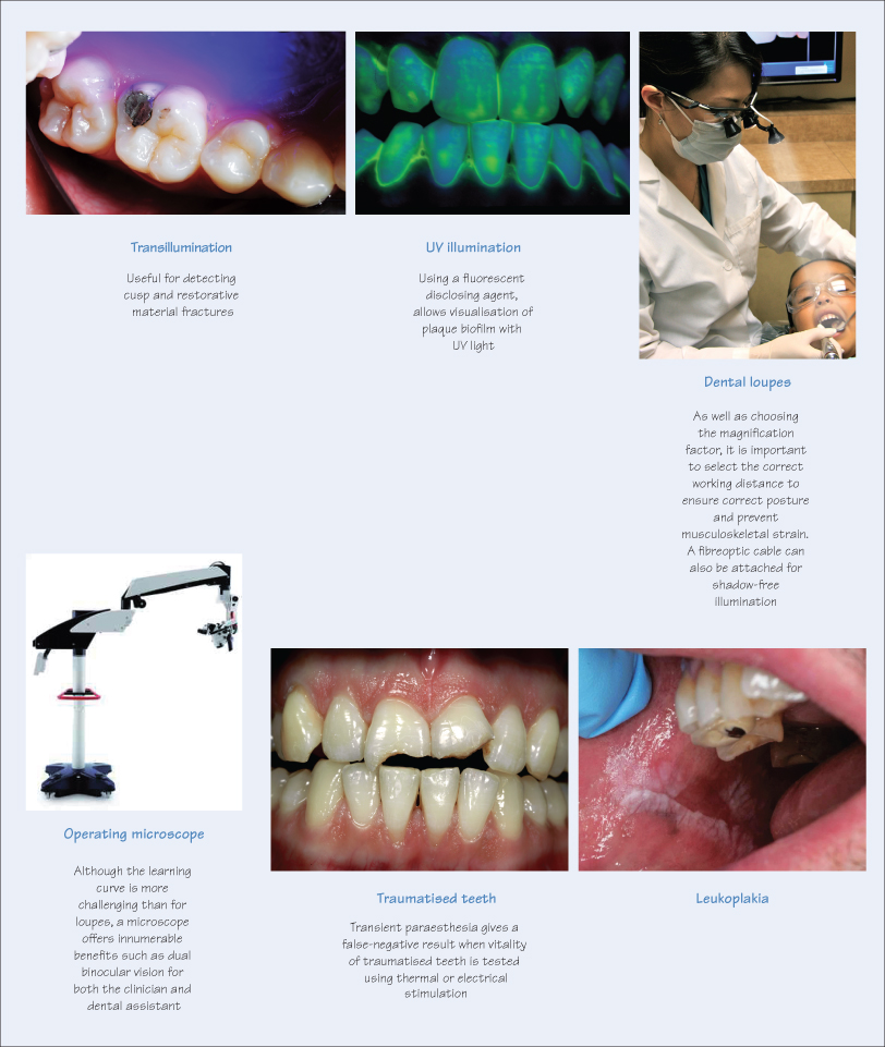
The typical illumination in a dental practice is the operatory light which delivers incident light that is suitable for most examinations. However, other types of illumination allow visualisation of features that may be missed by traditional light sources, e.g. transillumination via a fibreoptic cable emphasises fractures (tooth and restorations), defective restorative margins and caries. In addition, ultraviolet (UV) illumination is useful for detecting plaque biofilm (with fluorescent disclosing solutions) or porosity in ceramic restorations.
Magnification
Visual enhancement is not limited to specific specialties such as endodontics, but is invaluable in many dental disciplines such as prosthodontics and periodontics, and in detecting lesions that may be ‘invisible’ to the unaided eye. Magnification elevates visual acuity using loupes, intra-oral cameras, operating microscopes and projection stereomicroscopes using 3-D video technology. Loupes offer magnification from ×2 to ×5, while microscopes have the capability for ×20 or greater levels of reproduction.
The advantages of magnification are:
- Enhanced visual access to detail;
- Improved precision;
- Increased efficacy;
- Compensate for presbyopia;
- Comfortable ergonomics, avoiding musculoskeletal injuries.
The disadvantages of visual aids are:
- Limited field of view (beyond ×2.5);
- Reduced depth of field (with extreme magnifications >×10);
- Cross-infection concerns;
- Damage to optics (by dental debris, or air abrasion procedures);
- Learning curve (especially with microscopes).
Oral Precancer and Cancer Lesi/>
Stay updated, free dental videos. Join our Telegram channel

VIDEdental - Online dental courses


