Chapter 4
Infections of the Oral Mucosa
Aim
This chapter reviews the common oral mucosal and facial infections.
Outcome
After reading this chapter you should be familiar with the presenting features, investigations and management strategy for commonly encountered oro-facial infections.
Introduction
Infections of the oral mucosa are fall into three broad categories.
-
bacterial
-
viral
-
fungal.
Some take short-term advantage of a compromised immune system and others become chronic problems. When presented with an infection in clinical practice, in addition to consideration of the infecting organism, it is important that the circumstances of the infection are taken into account. This allows local, systemic or environmental factors to be identified and managed. A principle of managing infections is that treatment should follow identification of the active organism and its sensitivity to antimicrobial therapy. Close liaison with the microbiologist will ensure that appropriate samples are taken and properly transported to the laboratory. Antimicrobial therapy is not required for every patient. In many cases attention to local factors, such as the removal of an abscessed tooth, is all that is needed.
Bacterial Infections
Bacterial infections of the face and oral mucosa usually present as acute problems. They often represent local alterations in the host-bacterial balance, changing a commensal into a pathogen. Bacterial conditions seen include:
-
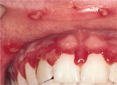
acute ulcerative gingivitis (Fig 4-1)
-
facial abscess
-
staphylococcal and streptococcal infections
-
bacterial mucositis
-
angular cheilitis (Fig 4-2)
-
impetigo
-
erysipelas
-
-
syphilis (Fig 4-3)
-
tuberculosis.
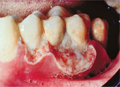
Fig 4-1 Acute necrotising ulcerative gingivitis (ANUG). The presentation of this condition warrants further investigation for possible underlying immunosuppression.
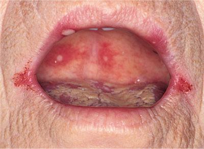
Fig 4-2 Aphthous type ulceration affecting the palate and tongue, with in addition bilateral angular cheilitis in a patient with vitamin B12 deficiency.
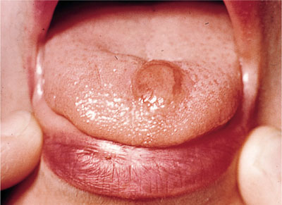
Fig 4-3 Primary syphilitic chancre on the dorsum of the tongue.
Acute Ulcerative Gingivitis
Acute ulcerative gingivitis is a necrotising condition of the marginal gingival tissues, seen mostly in smokers during the winter months. It is usually localised to the anterior gingivae and, as well as being painful, it also gives the patient a very unpleasant characteristic odour. In the immunocompromised the lesions may be found in any part of the dental arch and be much more extensive. In extreme cases they extend to include bone as well as soft-tissue necrosis. The cause is an anaerobic fuso-spirocheatal complex that is sensitive to metronidazole (200mg three times daily for seven days). Oral hygiene measures and topical use of chlorhexidine are often all that are required in the less severe cases in an immunocompetent host.
Facial Abscess
Facial abscesses are most commonly of dental origin and present with a swelling over the body or angle of the mandible or the infra-orbital region in patients who typically have dental neglect. If untreated, the infection will discharge onto the facial skin, producing an unsightly appearance and a persisting facial scar. The pattern of the discharge from a dental abscess to the skin or the mouth will depend on the position of the tooth roots relative to muscle insertions. Gravity then works to make most swellings from the upper teeth appear over the lower face and from the lower teeth either at the border of the mandible or the sublingual or submental area of the anterior neck. These are mixed infections of anaerobes and aerobes and require removal of the cause (extraction or pulpectomy) and, following culture and sensitivity assessment of a drained pus sample, a course of antibiotics may be provided to augment surgical management. Antibiotic therapy is not an alternative but is a supportive therapy. On rare occasions culture will suggest actinomycosis. Actinomyces species are common commensals in the mouth, but a true infection is uncommon.
Streptococcal and Staphylococcal Infections
Mucositis
Staphylococci are occasionally implicated in mucosal infection, particularly in the palate beneath dentures. The erythematous change is not clinically distinguishable from that produced by a chronic candidal infection and reinforces the need for microbiological investigation as part of the process used to reduce denture-induced stomatitis. Denture and oral hygiene measures are usually all that are required to settle the problem, which is mostly asymptomatic. Systemic antibiotics are rarely used.
Angular Cheilitis
This is a multifactorial problem. Systemic factors, such as iron deficiency or diabetes mellitus, act with local factors, such as a reduction in the facial vertical height due to a worn prosthesis, to allow staphylococci and candidal organisms to become established and pathogenic at the corners of the mouth. Management should address all issues, including correction of systemic factors, facial height and appliance hygiene. Often no more is required, but when the problem persists the use of an agent with both antifungal and antibacterial actions, such as miconazole, is preferred. Where the skin inflammation has been long-standing, the use of miconazole with hydrocortisone cream allows the skin architecture to return to normal by eliminating persisting inflammation. Abnormal skin is rapidly recolonised by the pathogens, and the angular cheilitis returns quickly.
Perioral Skin Infections (Fig 4-4)
Perioral skin infections are common in children and people who are immunocompromised. Impetigo is caused by Staphylococcus aureus, beta haemolytic streptococci or both. The staphylococci tend to produce thin-walled blisters, whereas a more crusting form tends to be associated with streptococci. Multiple lesions may present around the face but each area of impetigo remains discrete. It can spread rapidly to other individuals in close contact with the patient. Often the appearance can suggest recurrent herpes simplex to those unfamiliar with impetigo, and it is entirely possible that the skin breach initiated by herpes simplex may allow the entry of the impetigo organisms in some cases. Although a rare complication, an acute glomerulonephiritis may follow, in common with other streptococcal infections. Erysipelas is streptococcal in origin and the organism usually gains entry through an existing skin breach. It presents as a spreading erythematous facial infection with a well-defined advancing edge. The patient is usually systemically unwell and responds rapidly to penicillin treatment.
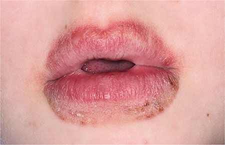
Fig 4-4 Lick eczema. Note how the area of inflammation extends as far as the tongue can reach. Occasionally this condition can become secondarily infected with Staphylococcous aureus to produce golden-coloured crusting.
Syphilis
Although in decline for many years, syphilis and its oral manifestations are again becoming more common. The infecting organism is Tremonema pallidum, and there are oral features in all three stages. These are:
-
the primary chancre
-
‘snail-track’ ulcers of the secondary stage
-
the gumma or leukoplakia of the tertiary stage.
Few practitioners have seen this condition nowadays, which makes diagnosis difficult. A high degree of suspicion and good laboratory collaboration is essential.
Tuberculosis
As with syphilis, the incidence of tuberculosis is on the rise, particularly in the immunosuppressed and immigrant populations. The common causative organism is Mycobacterium tuberculosis. In the immunocompromised host atypical mycobacteria may be responsible for infections, and it has been suggested that Mycobacterium paratuberculosis may be involved in Crohn’s disease and orofacial granulomatosis. In patients with active tuberculosis oral involvement is rare, but the organisms can produce enlarged cervical lymph nodes, an oropharyngeal abscess or a chronic ulcer on the tongue. Biopsy of a suspected lesion is essential to confirm the diagnosis, finding the tubercle bacilli in a chronic granulomatous reaction. Identification of a patient with tuberculosis requires notification to the public health services to identify, trace and screen any contacts of the patient and thus limit spread of the disease.
Viral Infections (Figs 4-5 to 4-8)
In the orofacial region, viral infections, particularly with the herpes group of viruses, are very frequent. The clinical problems encountered are given in Table 4-1.
Stay updated, free dental videos. Join our Telegram channel
VIDEdental - Online dental courses


