38 White lesions: Keratosis, leukoplakia
Figure 38.1 Biting causing keratosis.
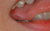
Figure 38.2 Biting mucosa causing keratosis.
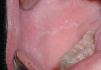
Figure 38.3 Cheek chewing.
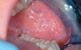
Figure 38.4 Frictional keratosis.
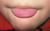
Figure 38.5 Leukoplakia etiology.

Figure 38.6 Leukoplakia: infective causes.
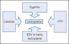
Figure 38.7 Homogeneous leukoplakia.
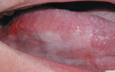
Figure 38.8 Verrucous leukoplakia.
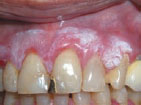
Figure 38.9 Leukoplakia that proved to be carcinoma.
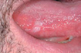
Figure 38.10a Acanthosis and hyperparakeratosis.
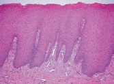
Figure 38.10b Keratosis and atrophy.

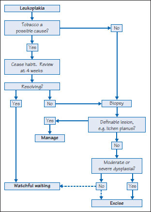
Figure 38.11 Leukoplakia management.
Definition: White lesion caused by repeated trauma.
Prevalence (approximate): Common.
Age mainly affected: Middle-age and older.
Gender mainly affected: M > F.
Etiopathogenesis: Etiological factors include prolonged abrasion (e.g. sharp tooth, dental appliance, toothbrushing, mastication, cheek biting). Bilateral alveolar ridge keratosis (BARK) may be seen in edentulous areas. An occlusal line (linea alba) is often seen on the lateral tongue (Figure 38.1) and in the buccal mucosae (Figure 38.2), as is cheek-biting (morsicatio buccarum or morsicatio mucosa oris, MMO), most prevalent in anxious females (Figure 38.3). Rarely self-mutilation is seen in psychiatric disorders (Figure 38.4), learning impairment or some rare syndromes.
Diagnostic features
Clinical features
Linea alba is typically thin, white with occasional petechiae and may be seen in isolation or />
Stay updated, free dental videos. Join our Telegram channel
VIDEdental - Online dental courses


