36 Ulcers and erosions: Erythema multiforme, toxic epidermal necrolysis and Stevens-Johnson syndrome
Figure 36.1 Erythema multiforme etiology.
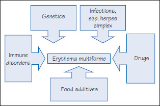
Figure 36.2 Erythema multiforme pathogenesis.
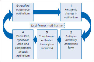
Figure 36.3 Erythema multiforme.
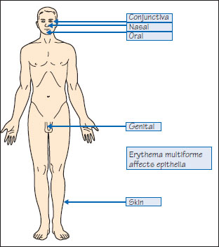
Figure 36.4 Erythema multiforme.
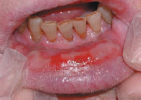
Figure 36.5 Erythema multiforme target lesions.
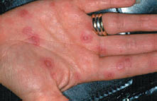
Figure 36.6 Erythema multiforme treatment.
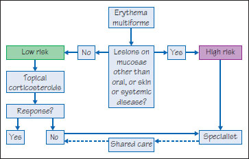
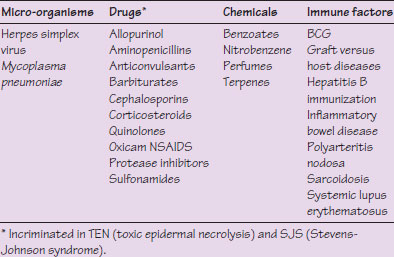
Table 36.1 Main causal factors in erythema multiforme.
Erythema multiforme
Definition: Erythema multiforme (EM) is a mucocutaneous condition mediated by antigen-antibody (immune complex – mainly IgM) deposition in the superficial microvasculature of skin and mucous membranes.
Prevalence (approximate): Uncommon.
Age mainly affected: Younger adults in second and third decades.
Gender mainly affected: M > F.
Etiopathogenesis: There may be a genetic predisposition, with various HLA associations (e.g. patients with extensive mucosal involvement may have HLA-DQB1*0402). A putative immunological hypersensitivity reaction usually to various micro-organisms or drugs (Figure 36.1) (Table 36.1), results in immune complexes and the ingress of cytotoxic CD T lymphocytes, inducing keratinocyte apoptosis and satellite cell necrosis (Figure 36.2).
Diagnostic features
EM minor (accounts for 80%) is a mild, self-limiting rash usually affecting one mucosa. EM major is a severe, life-threatening variant that overlaps with toxic epidermal necrolysis (see below) and involves m/>
Stay updated, free dental videos. Join our Telegram channel
VIDEdental - Online dental courses


