Moisture Control
Learning Outcomes
On completion of this chapter, the student will be able to achieve the following objectives:
• Pronounce, define, and spell the Key Terms.
• List isolation techniques used to decrease moisture during a dental procedure.
• Describe the two types of oral evacuation systems used in dentistry.
• Describe the grasp and positioning of the high-volume evacuator tip.
• Discuss the use of the air-water syringe.
• Describe the dental dam and its role in moisture control.
• List the equipment and supplies required for dental dam application.
• Describe the special preparation and placement of the dental dam.
Performance Outcomes
On completion of this chapter, the student will be able to meet competency standards in the following skills:
• Perform the grasp and positioning of the high-volume evacuator during a procedure.
• Perform limited-area and full-mouth rinses.
• Place cotton rolls for isolation.
• Identify the equipment and supplies used for dental dam application.
• Have the dental dam prepared correctly for a procedure.
Electronic Resources
 Additional information related to content in Chapter 36 can be found on the companion Evolve Web site.
 and the Multimedia Procedures DVD
Key Terms
Aspirate (AS-pi-rayt) To draw back or to draw within.
Bow Rounded part of clamp that extends through the dental dam.
Exposed Pertaining to selected teeth visible through the dam; isolated.
Invert (in-VURT) To reverse the position, order, or condition. To turn inside out or upside down.
Isolated Pertaining to selected teeth visible through the dam; exposed.
Jaws Part of a clamp that is shaped into four prongs to help stabilize the clamp on the tooth.
Malaligned (mal-uh-LINED) Displaced out of line, especially teeth displaced from normal relation to the line of the dental arch; also called malposed.
Septum (SEP-tum) Dental dam material located between the holes of the punched dam.
Stylus (STYE-lus) Sharp, pointed tool used for cutting.
Universal Pertaining to the same clamp that can be placed on the same type of tooth in the opposite quadrant.
Winged clamp Type of dental dam clamp that has extensions to help retain the dental dam.
When assisting in a dental procedure, one of the most important responsibilities of the chairside dental assistant is to maintain moisture control. The assistant keeps the operating field free of excess water, saliva, blood, tooth fragments, and excess dental materials.
This chapter describes several moisture control techniques and their incorporation into the dental procedure.
Oral Evacuation Systems
The term oral evacuation describes the process of removing excess fluid and debris from the mouth. The use of an oral evacuator completes this process before, during, and after a dental procedure. The two types of evacuators most often used in a dental procedure are the saliva ejector and the high-volume evacuator (HVE).
Saliva Ejector
The saliva ejector is a small, straw-shaped oral evacuator that is used during less-invasive dental procedures (Fig. 36-1). The main function of the saliva ejector is to remove liquids from the mouth; it is not powerful enough to remove solid debris. Indications for using the saliva injector include the following:
• Preventive procedures, such as prophylaxis, placement of sealants, and fluoride treatments
• Control of saliva and moisture accumulation under the dental dam
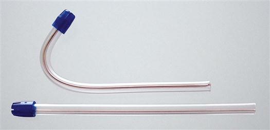
The saliva ejector is made of a soft plastic tubing that can be shaped for easy placement in the oral cavity. The ejector can be held (1) throughout the procedure, with the use of repeated sweeps of the mouth to remove fluids, or (2) by positioning the suction tip in the mouth during a procedure.
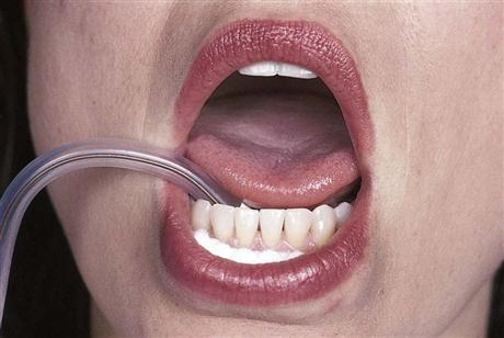
To place the saliva ejector in a stationary position, bend and form the tubing into the shape of a candy cane (Fig. 36-2). This shape allows the saliva ejector to be positioned under the tongue, where most fluids accumulate. When placing the saliva ejector, insert a cotton roll to act as a buffer, so that it will not traumatize oral tissues. When positioning the saliva ejector for a procedure, position it on the opposite side from where the dentist is working to reduce the number of items in the operating field.
High-Volume Evacuator
The HVE is used to remove saliva, blood, water, and debris during a dental procedure. The HVE system, also known as the oral evacuator, works on a vacuum principle similar to that of a household vacuum cleaner. It is able to take up water and debris because of the high volume of air that is moved into the vacuum hose at low pressure to create the strong suction. The HVE has three main purposes:
• To keep the mouth free of saliva, blood, water, and debris
• To retract the tongue and cheek away from the field of operation
• To reduce the bacterial aerosol caused by the high-speed handpiece
Suction Tips
Oral evacuation tips for the HVE system are designed to accommodate different types of procedures.
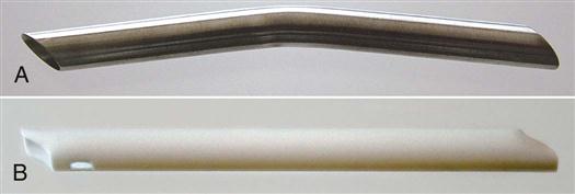
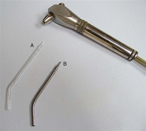
Operative suction tips are larger in circumference and designed with a straight or slight angle in the middle. Each opening has a beveled (having a surface angle that meets another angle) working end, so that the tip can be positioned parallel to the site for better suction. Most HVE tips are made of a durable plastic and are disposed of after a single use, or stainless steel, which must be sterilized before reuse (Fig. 36-3).
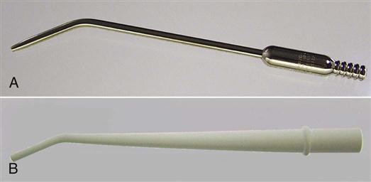
Surgical suction tips are much smaller in circumference. This design is critical to placement within the surgical site, which typically is more limited in size and visibility. The concern in a surgical procedure is the removal of blood, tissue, and debris, rather than a large amount of water and fluid. Surgical tips are made of stainless steel and are part of the surgical setup (Fig. 36-4). Refer to Chapter 56 for further discussion.
Grasping the Evacuator
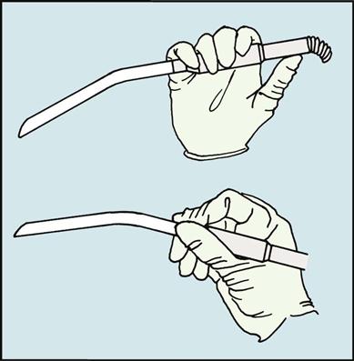
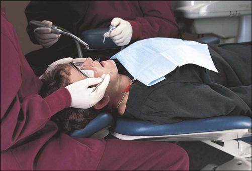
The HVE may be held in the thumb-to-nose grasp or the pen grasp (Fig. 36-5). Either method provides the dental assistant with control of the tip, which is necessary for patient comfort and safety.
Positioning the Evacuator
Depending on the resistance of the tissue to be retracted and the area being treated, you may wish to alternate between ways of positioning the evacuator tip. When assisting a right-handed dentist, the assistant grasps the evacuator in the right hand. When assisting a left-handed dentist, the assistant grasps the evacuator in the left hand. The hand that is not holding the suction is free to operate the air-water syringe or to participate in the transfer of instruments to the dentist as needed. HVE positions discussed in this chapter are for use with a right-handed dentist.
As a dental assistant, you must understand your role in placing and operating the oral evacuation system. The timing of when suction is used, when the HVE is positioned, and when suction is removed from the mouth is critical to the efficient and effective performance of the procedure. Specific guidelines for positioning the HVE include the following (Procedure 36-1):
• Place the evacuator before the dentist positions the handpiece and the mouth mirror.
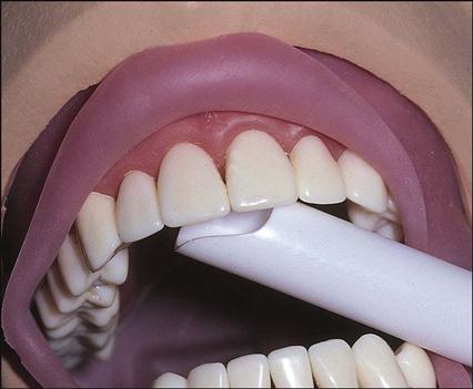
• Position the suction tip on the surface of the operative tooth closest to you (Fig. 36-6).
• Position the bevel of the suction tip so that it is parallel to the tooth surface.
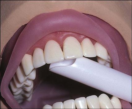
• Keep the edge of the suction tip even with or slightly beyond the occlusal surface or the incisal edge (Fig. 36-7).
PROCEDURE 36-1 


Positioning the High-Volume Evacuator During a Procedure
Equipment and Supplies
Procedural Steps
1 Place the HVE tip in the holder by pushing the end of the tip into the holder through the plastic barrier.
Purpose: Leaves the opposite end exposed and ready for use.
2 If necessary, use the HVE tip or a mouth mirror to gently retract the cheek or tongue.
Posterior Placement
3 For a mandibular site, place a cotton roll under the suction tip.
Purpose: Provides patient comfort, aids in stabilizing tip placement, and prevents injury to the tissues.
4 Place the bevel of the HVE tip as close as possible to the tooth that is being prepared.
Purpose: Suction will draw the water into the tip immediately after it leaves the tooth that is being prepared.
6 Place the upper edge of the HVE tip so that it extends slightly beyond the occlusal surface.
Purpose: Suction will catch the water spray from the handpiece as it leaves the tooth that is being prepared.
Anterior Placement
Daily Maintenance of the Evacuation System
As with any piece of equipment, the HVE must undergo daily upkeep to maintain proper working order. Specific maintenance guidelines for the evacuation systems and the air-water syringe include the following:

Rinsing the Oral Cavity
Frequent rinsing of the oral cavity maintains a clear operating field for the dentist and keeps the patient comfortable. Rinsing also removes debris from the patient’s mouth before dismissal. The two basic types of rinsing procedures used in dentistry are limited-area rinsing and complete mouth rinsing, also referred to as a full-mouth rinse.
Limited-Area Rinsing
This is performed frequently throughout a procedure as debris accumulates during the preparation and restoration of a tooth. This rinsing must be accomplished quickly and efficiently, causing no delay in the procedure. Limited-area rinsing is frequently accomplished when the dentist exits the mouth and pauses for inspection.
Full-Mouth Rinsing
The full-mouth rinse is used when the patient’s entire mouth needs freshening. Complete mouth rinsing may be completed after a long restorative procedure, after dental prophylaxis, or before patient dismissal following any dental procedure.
The saliva ejector can be used as an alternative to the HVE in the complete mouth rinse procedure when the assistant performs this alone.
Air-Water Syringe
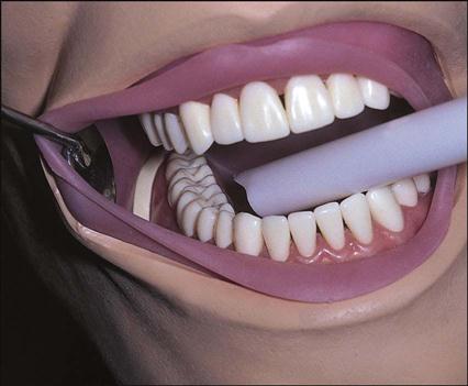
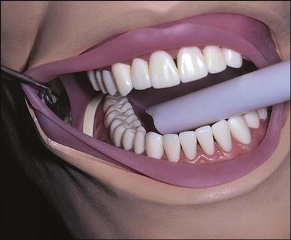
To complete the rinsing process, the air-water syringe is used for convenience and accuracy. As described in Chapter 32, the air-water syringe is connected to the dental unit and provides air, water, or a combination through a small sterile tip. The attached disposable tip can be positioned for the maxillary and mandibular arches by directing the end toward the specific arch (Fig. 36-8). Guidelines for using the air-water syringe include the following (Procedure 36-2):
• Direct the syringe tip toward the tooth that is being treated.
• Use the air on the mouth mirror continuously when indirect vision is involved.
• When you hear the handpiece stop, rinse and dry the site.
• When completing a limited-area or full-mouth rinse, move the tip while spraying the area.

PROCEDURE 36-2 

Performing a Mouth Rinse
Equipment and Supplies
Procedural Steps
1 Decide which oral evacuation system would be best for use in the rinsing procedure.
2 Grasp the air-water syringe in your left hand and the HVE or saliva ejector in your right hand.
Limited-Area Mouth Rinse
3 Turn on the suction, and position the tip toward the site for a limited-area rinse.
4 Spray the combination of air and water onto the site to be rinsed.
Purpose: The combination of air and water provides greater force to clean the area thoroughly.
5 Suction all fluid and debris from the area, making sure to remove all fluids.
Full-Mouth Rinse
7 Have the patient turn toward you.
Purpose: Turning the head allows the water to pool on one side, making it easier for you to suction.
8 Turn on the HVE or saliva ejector, and position it in the vestibule of the patient’s left side.
Note: Position the tip carefully so that it does not come into contact with soft tissue.
Stay updated, free dental videos. Join our Telegram channel
VIDEdental - Online dental courses


