32 Poor quality first permanent molars
Examination
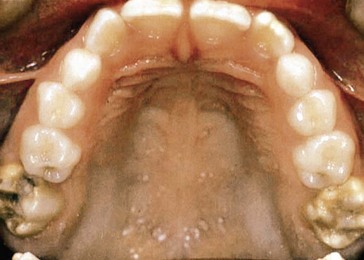
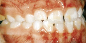
Intraoral examination revealed that all four first permanent molars (FPMs) were hypomineralized with areas of brown, rough enamel. In addition there were areas of hypoplasia where the enamel had chipped away, exposing the underlying dentine. The maxillary molars were the worst affected by hypoplasia.  had a large amalgam restoration. In addition some white and brown hypomineralized areas were visible on the labial surfaces of the newly erupted upper and lower permanent central incisors (Figs 32.1 and 32.2).
 had a large amalgam restoration. In addition some white and brown hypomineralized areas were visible on the labial surfaces of the newly erupted upper and lower permanent central incisors (Figs 32.1 and 32.2).
Upper and lower arches were uncrowded. Space assessed from the distal of 2’s to mesial of 6’s in each quadrant was 21.5 mm in the lower arch and 22 mm in each upper arch quadrant. On average, 21 mm and 22 mm are required for these distances in the lower and upper arches, respectively.
 Do you think that the enamel hypomineralization and hypoplasia noted on the first permanent molars and the permanent incisors follows a chronological pattern? If so, at what time was the affected enamel formed?
Yes, it is possible that there is a chronological pattern. The cusp tips of the first permanent molars begin mineralizing from the eighth month of pregnancy. The cusp tips of the incisors and cuspids (canines) from about 3 months of age (the upper lateral incisor slightly later at 10–12 months). Mineralization dates for the permanent dentition are given in Table 32.1.
| Tooth | Mineralization begins (months) |
|---|---|
| Upper | |
| Central incisor | 3–4 |
| Lateral incisor | 10–12 |
| Canine | 4–5 |
| First premolar | 18–21 |
| Second premolar | 24–27 |
| First molar | At birth |
| Second molar | 30–36 |
| Third molar | 84–108 |
| Lower | |
| Central incisor | 3–4 |
| Lateral incisor | 3–4 |
| Canine | 4–5 |
| First premolar | 21–24 |
| Second premolar | 27–30 |
| First molar | At birth |
| Second molar | 30–36 |
| Third molar | 96–120 |
Stay updated, free dental videos. Join our Telegram channel

VIDEdental - Online dental courses


 ; the molar relationship was Class I bilaterally.


