31
Impacted teeth
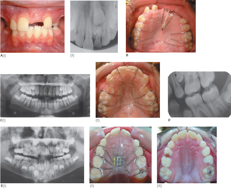
Figure 31.1 (A) (i) An asymmetrical pattern of central incisor eruption and eruption of the lateral incisors before a central incisor indicates a problem with central incisor eruption, (ii) Further investigation shows that the upper left central incisor has been prevented from erupting by multiple supernumerary teeth. (B) (i) An example of an upper removable appliance (URA) being used to align impacted central incisors. The incisors have been exposed and bonded with a gold chain. Elastic traction is applied to the unerupted teeth from the URA via the gold chain. (C) (i) Early loss of second deciduous molars has resulted in maxillary second premolar impaction, (ii) Extraction of the first premolars, followed by space maintenance with a URA has resulted in the eruption of the second premolars into a good position. Surgical removal of the second premolars has been avoided. (D) The first premolars should only be extracted to create epace if the second premolars are of normal form. In this case, radiographic examination revealed that the crowne of the second premolars were hypoplastic. The second premolars should be extracted in such cases. (E) (i) The upper left first permanent molar (UL6) is impacted and causing root resorption of the upper left second deciduous molar, (ii) URA used to apply a distalising force via an occlusal button bonded onto UL6. (iii) Following correction of UL6.
Impaction can be defined as the failure of a tooth to erupt, usually due to crowding or an obstruction (e.g. supernumerary tooth, fibrous tissue). Rarely a tooth may fail to erupt due to failure in the eruptive mechanism (primary failure of eruption, Chapter 9). Some medical conditions (e.g. cleidocranial dysplasia, Table 13.4iii) can be associated with multiple impactions. The aim of this chapter is to give a brief overview of impacted teeth. Chapter 32 focuses on maxillary canine impaction.
Impacted maxillary central incisors
The prevalence of impacted maxillary central incisors is reported to be 0.13%. The most common aetiological factor is the presence of supernumerary teeth but other factors such as trauma (e.g. dilaceration), retention of primary incisors and impaction against fibrous tissue may be important. Impaction should be suspected if the contralateral incisor has erupted more than six months previously or if the lateral incisors have erupted before the central incisors (Figure 31.1A). In such cases a radiographic assessment using an upper anterior occlusal or periapical view(s) is recommended.
The first principle of management is to ensure that adequate space is present to accommodate the impacted tooth (9 mm for an average incisor). This may involve extraction of retained deciduous teeth. Secondly, any cause of obstruction should be removed. Tuberculate supernumeraries are more commonly associated with incisor impaction than conically shaped teeth (see Chapter 34). In up to 75% of cases, the impacted incisor may erupt spontaneously within 16 months of removal of the supernumerary. Spontaneous improve/>
Stay updated, free dental videos. Join our Telegram channel
VIDEdental - Online dental courses


