The temporomandibular joint
Introduction
From the investigative point of view the knowledge required by clinicians includes:
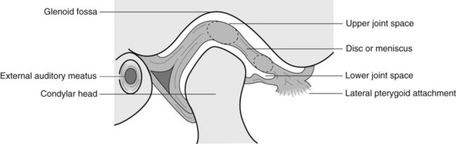
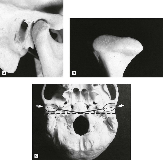
Normal anatomy
The basic components of the TMJ include:
• The mandibular component, i.e. the head of the condyle
• The temporal component, i.e. the glenoid fossa and articular eminence
• The capsule surrounding the joint (see Figs 30.1 and 30.2).
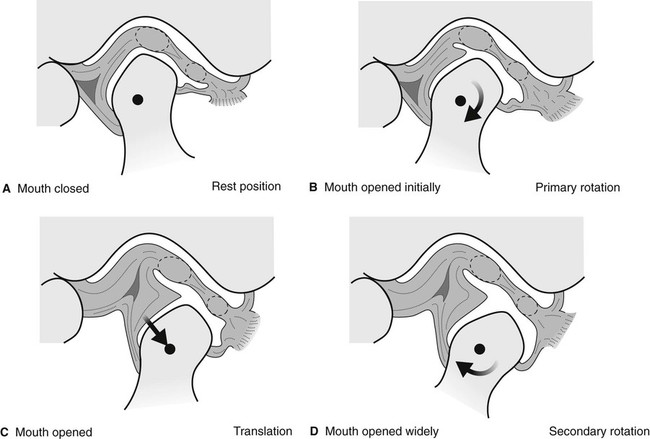
In addition to this knowledge of the static anatomy, clinicians need to be aware of the types and range of joint movements which result in the condyles moving downwards and forwards when patients open their mouths. These include:
Investigations
Modern imaging of the TMJ is dependent on the facilities available but could include:
• Conventional panoramic radiography
• Specific field limitation TMJ panoramic programmes
Previously described transorbital and transcranial views are now seldom used and are only of historical interest.
Panoramic radiography
Main indications
The main clinical indications include:
• TMJ pain dysfunction syndrome if bone anomalies are suspected clinically
• To investigate disease within the joint
• To investigate pathological conditions affecting the condylar heads
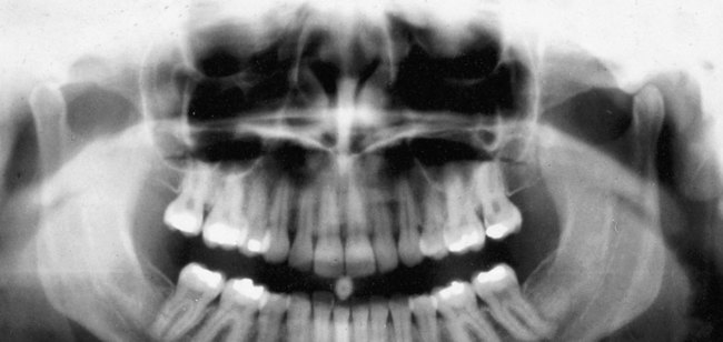
Panoramic TMJ programmes
Main indications
Technique summary
The technique can be summarized as follows:
• The patient is positioned with their Frankfurt plane angled 5° downwards within a panoramic unit with their mouth closed but using a special nose/chin support as shown in Fig. 30.5A instead of the bite-peg
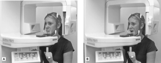
• The head is accurately positioned using the light beam markers and immobilized using the temple supports
• The distance from the external auditory meatus to the canine light is measured and the anteroposterior position of the chin support adjusted manually to ensure that the condyles appear in the middle of the image
• During the exposure, first the left and then the right condyle is imaged in the closed position
• The equipment automatically returns to the start position
• The patient is instructed to open the mouth, as shown in Fig. 30.5B
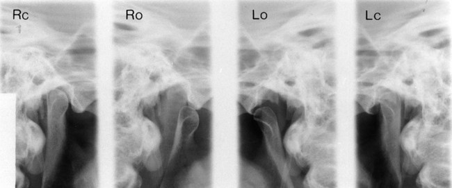
• The left and right condyles are then exposed in the open position and the resultant image is shown in Fig. 30.6.
Transpharyngeal radiography
Main indications
The main clinical indications include:
• TMJ pain dysfunction syndrome
• To investigate the presence of joint disease, particularly osteoarthritis and rheumatoid arthritis
• To investigate pathological conditions affecting the condylar head, including cysts or tumours
Technique and positioning
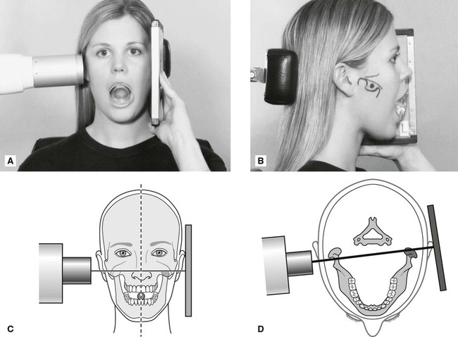
1. The patient holds the cassette against the side of the face over the TMJ of interest. The film and the mid-sagittal plane of the head are parallel. The patient’s mouth is open and a bite-block is inserted for stability.
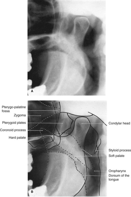
2. The X-ray tubehead is positioned in front of the opposite condyle and beneath the zygomatic arch. It is aimed through the sigmoid notch, slightly posteriorly, across the pharynx at the condyle under investigation, as shown in Fig. 30.7. Usually this view is taken of both condyles to allow comparison.
Stay updated, free dental videos. Join our Telegram channel
VIDEdental - Online dental courses


