Variations in Transition of Mandibular Posterior Teeth*
The space available in the dental arch for transition of the posterior teeth is bounded by the distal surface of the lateral permanent incisor crown and the mesial surface of the first permanent molar crown. The sum of the mesiodistal crown dimensions of the mandibular deciduous molars and canine exceeds that of their successors. The first premolar usually has a mesiodistal crown width similar to that of its predecessor or slightly smaller. The second premolar has a considerably smaller mesiodistal crown dimension than its predecessor. The reverse situation is common for the canines. These differences in mesiodistal crown dimensions of the corresponding deciduous and permanent posterior teeth are responsible for some typical features of the transition.
Distinct differences exist between the two jaws regarding the transition of the posterior teeth. These differences are associated with the relatively large middle section of the apical area in the mandible and the small one in the maxilla. Mandibular permanent canines and premolars erupt from spacious conditions in the middle section of the apical area to more limited ones in the dental arch. In the maxilla the space for the crowns of the corresponding teeth increases when they move from the middle section of the apical area to the dental arch.
The transition of the mandibular posterior teeth is discussed in relation to the space available for the permanent canine and premolars in the middle section of the apical area and in the dental arch. The variation in location of the permanent canine and the premolars and their relation to their predecessors and the adjacent lateral permanent incisor and first molar are indicated. The differences in emergence sequence and associated preemergence and postemergence behaviour are discussed. Nine transition patterns are distinguished, five of which are presented in detail.
The anterior section of the apical area is bounded laterally by the mesial surfaces of the permanent canines (see The anterior section of the apical area). The dimensions in the deciduous dental arch are of limited relevance for the size of the anterior section of the apical area, because the correlation between the mesiodistal crown dimensions of the corresponding deciduous and permanent anterior teeth is rather low. The space that is initially available in the dental arch is usually less than that required for the arrangement of the four mandibular permanent incisors in good alignment. During the transitional process the space available will increase as the canines move distally and laterally.
The situation is different for the canine-premolar region, where normally the interdental arch dimension from the distal surface of the crown of the lateral permanent incisor to the mesial surface of the first permanent molar does not increase but decreases with the transition of the posterior teeth. This dimension is slightly larger than the combined mesiodistal crown widths of the permanent teeth that emerge there. A lack in overall space does not normally exist and the middle section of the apical area is accordingly adequate in size. However, problems may arise when some of the excess space is utilised for an improved alignment of the incisors and when the emergence sequence is unfavourable. Premature loss of deciduous tooth material due to decay or extraction may considerably influence the course of events and the result of the transition of the posterior teeth.
The size and shape of the middle section of the apical area vary on the basis of (1) the overall morphology of the mandible, (2) the location of the permanent canine within the jaw and its relation to the adjacent lateral permanent incisor, and (3) the position of the first permanent molar. The latter depends on the mesiodistal crown dimensions of the deciduous molars and the molar’s location during formation. Its subsequent mesiodistal position in the dental arch is mainly determined by its relation to the distal surface of the adjacent second deciduous molar.
Invariably the permanent canines are initially formed closer to the lower border of the mandible than the second premolars and particularly the first premolars. In the preeruption phase the forming parts of the premolars are closer to the level of the occlusal plane than of the canine, which becomes the longest tooth in the mandible. The tip of the canine crown is located lingual to the apex of its predecessor. However, the buccal aspect of its crown bulges out and is easily palpable. The premolars are formed between the roots of their predecessors. Prior to eruption the distal surface of the permanent canine crown is in contact with or in close proximity to the mesial side of the cementoenamel junction of the first premolar.
Considerable variation can exist in the relation between the permanent canine crown and the root of its predecessor, as has been discussed and demonstrated in Chapter 2. Also, the relation between the premolars and their predecessors can vary markedly, more so in the mesiodistal than buccolingual direction.
The size of the middle section of the apical area in relation to that of the anterior section depends partly on the positions of the permanent canine and adjacent lateral incisor and partly on the labiolingual inclination of the anterior alveolar process. As for the anterior region, large, medium, and small sections of the middle apical areas are distinguished (Figs. 3-1 to 3-3).
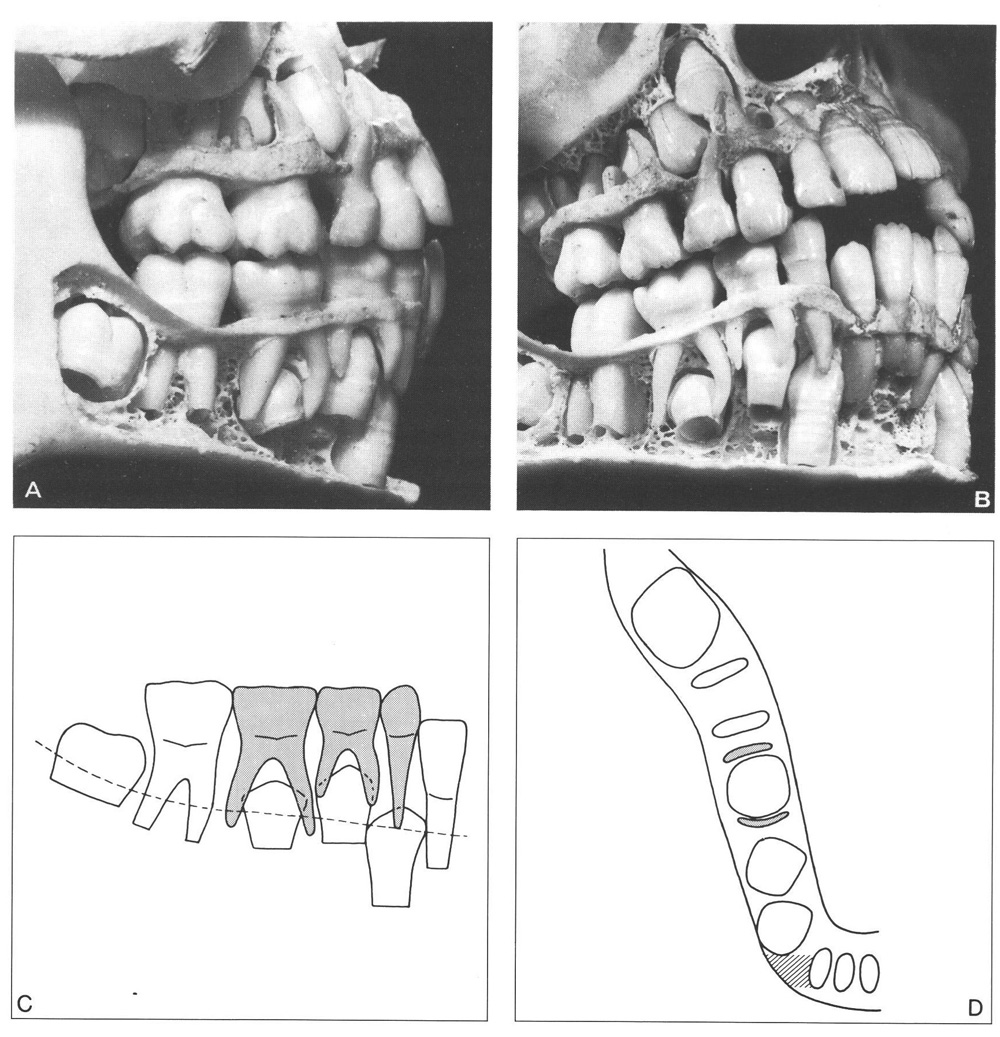
Fig. 3-1 Large middle section of the apical area in the mandible. The position of the permanent canine, the differences in mesiodistal crown dimensions of corresponding teeth, and the size and shape of the middle section of the apical area in the mesiodistal direction are favourable.
A The mesiodistal crown width of the mandibular second deciduous molar is considerably larger than that of its successor. The latter is at some distance from the mesial root of the first permanent molar. The mandibular permanent canine is only slightly mesially angulated.
B Between the crowns of the premolars is a space of about 3 mm. However, the mesial surface of the first premolar is in contact with the distal corner of the permanent canine crown. The latter is slightly distal to the apex of its predecessor and distal to the root of the lateral incisor.
C Schematic drawing with a dotted line indicating the section presented in D. Note the relation between the permanent canine crown and the root of the lateral incisor.
D Excess space is available between the mesial root of the first permanent molar and the distal surface of the root of the lateral incisor for the crowns of the premolars and permanent canine. Note the size of the indicated area between the permanent canine and the lateral incisor and particularly its sagittal dimension.
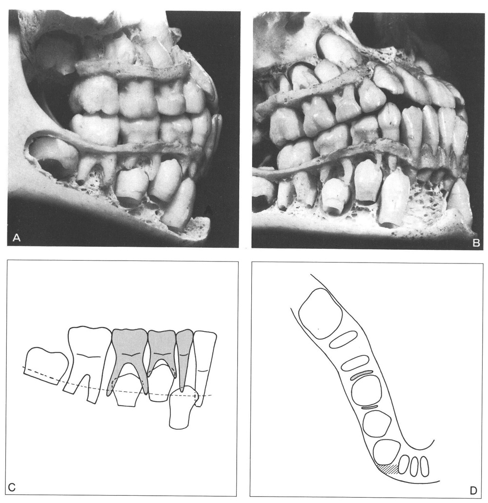
Fig. 3-2 Medium middle section of the apical area in the mandible. The position of the permanent canine, the differences in mesiodistal crown dimensions of the corresponding teeth, and the size and shape of the middle section of the apical area are critical.
A The mesiodistal crown width of the second deciduous molar is only slightly larger than that of its successor. The latter is rather close to the mesial root of the first permanent molar. The permanent canine is mesially angulated.
B Between the crowns of the premolars is a space of approximately 1 mm. The mesial surface of the first premolar is in close proximity to the distal surface of the permanent canine. The mesial side of the crown of the latter overlaps the root of the lateral incisor.
C The crown of the permanent canine is mesial to the apex of its predecessor. Note the relationship between the permanent canine crown and the lateral incisor root.
D The space available between the mesial root of the first permanent molar and the distal surface of the root of the lateral incisor is sufficient but critical. The crown of the permanent canine must move distally to establish a good contact with the crown of the lateral incisor. Extra space is required, and it is questionable if that extra space can be provided from the distal side. The size of the indicated area between the permanent canine crown and the root of the lateral incisor is critical, particularly in a sagittal direction.
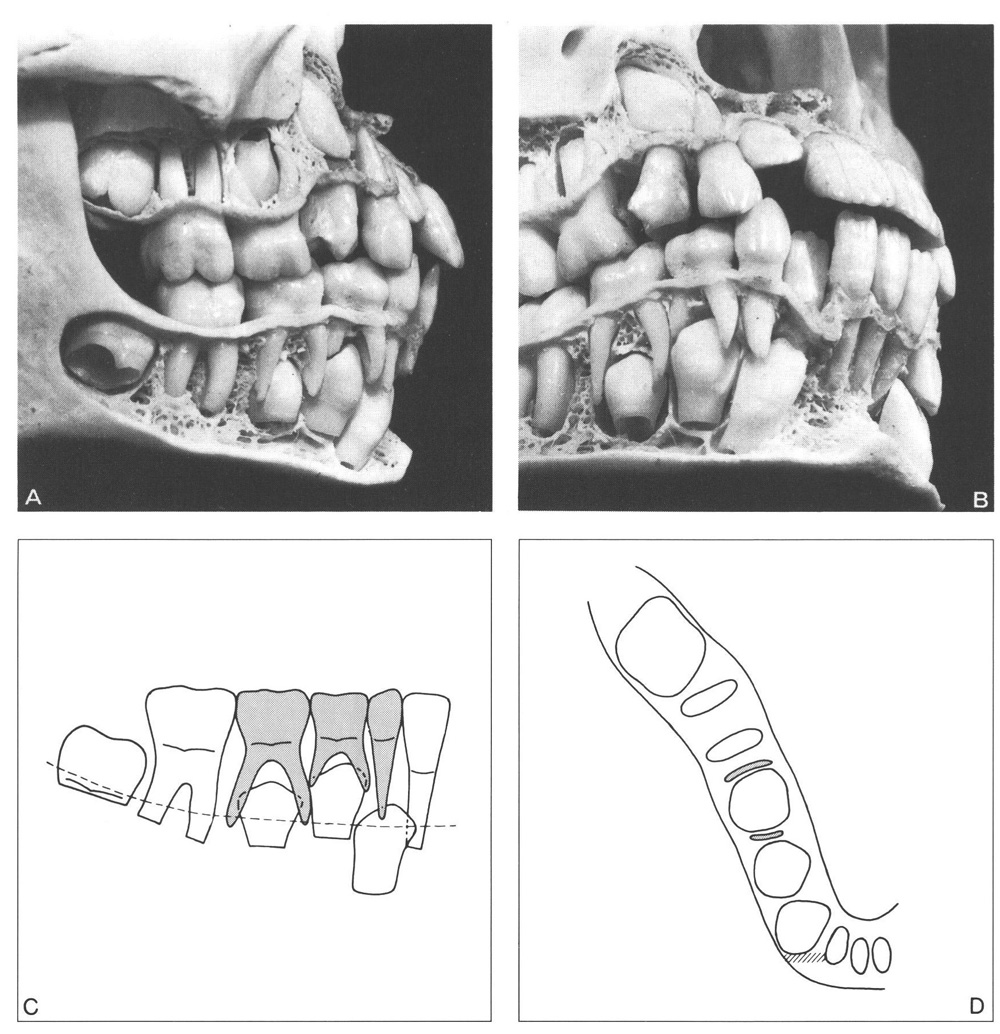
Fig. 3-3 Small middle section of the apical area in the mandible. The position of the permanent canine, its relation to the lateral incisor, the differences in mesiodistal crown dimensions of the corresponding teeth, and the size and shape of the middle section of the apical area are unfavourable.
A Some space exists between the distal surface of the crown of the second premolar and the mesial roots of the first permanent molar. However, the canine is distinctly mesially located and angulated.
B The differences in mesiodistal crown width of the canines and the first deciduous molar/premolar are unfavourable and overrule the favourable difference in that respect for the second deciduous molar/premolar. The crowns of the premolars are close together; the distal surface of the permanent canine crown lies approximately in the same plane as the distal surface of the root of its predecessor.
C The crowns of the permanent teeth that must still erupt are close together and crowded. The mesial surface of the permanent canine overlaps more than half of the root of the lateral incisor.
D To establish a harmonious contact between the crown of the lateral incisor and that of the permanent canine, the latter must move distally over a considerable distance, which is in fact impossible. The space available in the dental arch between the mesial root of the first permanent molar and the distal surface of the lateral incisor is too small to accommodate the permanent canine and premolars without crowding. The indicated area between the permanent lateral and permanent canine and particularly its sagittal dimension is a clear sign of the unfavourable conditions in that region.
The differences in the sum of the mesiodistal crown dimensions of the two deciduous molars and canine and that of their successors are crucial. A marked difference can therefore exist between the mesiodistal crown dimensions of deciduous molars and canines and that of their successors, particularly for the canines. The differences in crown width of the corresponding individual teeth together with the sequence of emergence play an important role in the transition process of the posterior teeth.
The sequence of emergence in the transition of the posterior teeth is not uniform as in the incisal region. The arrangement of the premolars and permanent canine within the jaw and their relation to the roots of the deciduous teeth and adjacent permanent teeth allow a variation in emergence sequence. The spatial conditions may also allow the two premolars and permanent canine to emerge simultaneously. Particularly in a large middle section of the apical area, one tooth does not need to emerge prior to its neighbour to create the space for the latter to erupt, as is typical for the incisor region. In most instances the mandibular permanent canine emerges first. In its preemergence eruption phase it passes the first premolar, which is initially closer to the occlusal plane. The canine has the longest distance to travel in its eruption; the first premolar the shortest. The second premolar, generally the last one to emerge, is initially located at a vertical level somewhere between those of the two other teeth. It therefore assumes an intermediate position regarding the length of the eruption path. The variation in initial direction of eruption and the subsequent eruption behaviour are greater in a large and also in a small middle section of the apical area than in a medium-sized area. It would appear that in a large middle section of the apical area the crowns have the freedom to be arranged in varying ways. In this situation the premolars can erupt in diverse directions. In a medium middle section of the apical area the canine and premolar crowns are situated in restricted spatial conditions, allowing less variation in their orientation. In a small middle section of the apical area the canine and premolar crowns are arranged in crowded conditions with the accompanying deviations in orientation.
In Fig. 3-4 the process of transition of the mandibular and maxillary posterior teeth is illustrated and explained for the most frequently observed sequences of emergence. In Fig. 3-5 some variations in the sequence of emergence of the posterior teeth, including the second permanent molar, are indicated. The moment of emergence of the latter in relation to the transition of the posterior teeth and particularly to the loss of the second deciduous molar is important with regard to the amount of mesial migration of the first permanent molar associated with the transition of the posterior teeth. The first permanent molar will become more mesially displaced when the second permanent molar emerges prior to the loss of the second deciduous molar than if it emerges after that event.222
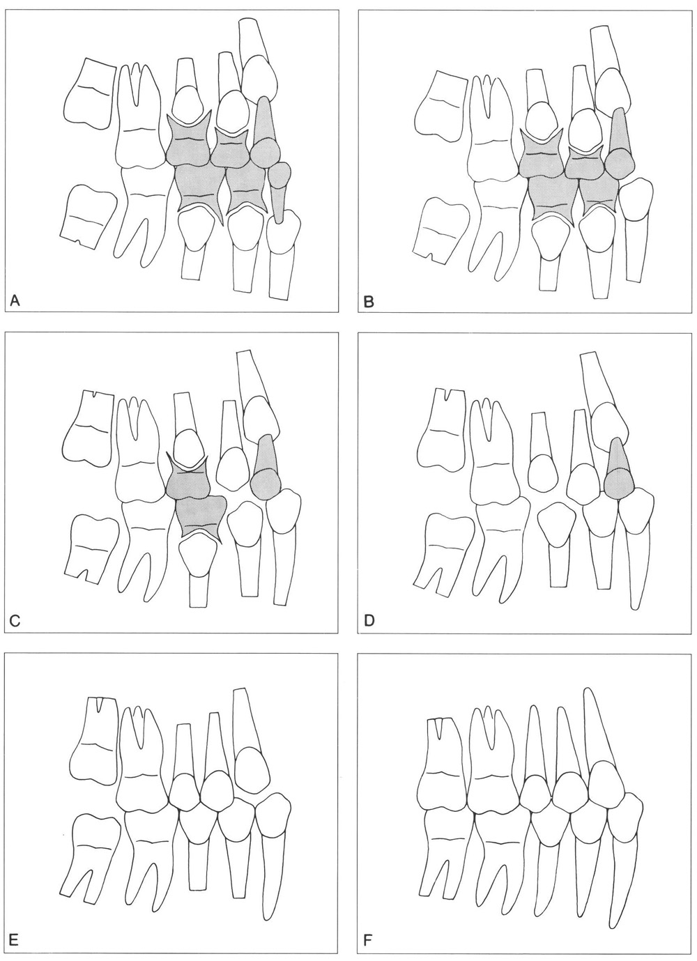
Figure 3-4
Fig. 3-4 Schematic presentation of the transition of the mandibular and maxillary posterior teeth and the emergence of the second permanent molars.
A The premolar crowns are located between the roots of their predecessors, which resorb concomitantly with the eruption of the premolars. In both jaws the occlusal surface of the permanent canine is initially at a greater distance from the occlusal plane than that of the first premolar. In this illustration the mandibular canine has already started to erupt and is at the same level as the first premolar. Both mandibular premolars are approximately perpendicularly oriented in relation to the occlusal plane. The permanent mandibular canine is slightly mesially angulated. In a large middle section of the apical area in the mandible some space may exist between the canine and first premolar crown. The mandibular permanent second molar is mesiolingually oriented in accordance with its initial position.
In the maxilla the first premolar is initially the closest to the occlusal plane and normally maintains that condition and emerges as the first one of the three teeth involved. The distal corner of the permanent canine crown is situated close to the mesial-cervical region of the first premolar. As in the mandible, the second premolar takes an intermediate position regarding the distance between the crown and the occlusal plane. Both maxillary premolars are slightly mesially angulated, the canine somewhat more. The maxillary permanent second molar is distobuccally oriented in accordance with its initial position and the local spatial conditions in the tuberosity region.
B The mandibular canine emerges shortly after the loss of its predecessor and has left the adjacent premolar behind. The diastema originally present between the deciduous canine and first deciduous molar provides the extra space required to accommodate the wider permanent canine crown within the dental arch. In the maxilla the first premolar has erupted more than the two adjacent teeth.
C The mandibular first premolar emerges shortly after the canine. Sufficient space is available. In the maxilla the first premolar also emerges with adequate space.
D The mandibular second premolar emerges into an excess of space in accordance with the differences in mesiodistal crown dimensions involved. In the maxilla a comparable situation is found, though with less excess space.
E The mandibular premolars are fully erupted and the excess space is reduced mainly by mesial migration of the mandibular first permanent molar. In the maxilla the canine emerges as the last of the three.
F The second permanent molars have emerged and attained occlusion. In the maxilla the permanent canine has taken up its position within the dental arch. The extra space required for the wide permanent crown was obtained from the remaining diastemata in the incisal region and the excess remaining after the emergence of the premolars. The maxillary second permanent molar has emerged and occludes with the mandibular molar.
On the average the process of transition of the posterior teeth only lasts about 1 to 1.5 years, but it may take considerably longer. The sequence of emergence can differ markedly and all theoretically possible sequences occur. The maxillary permanent canine usually does not emerge prior to the first premolar unless excess space is available in that region, or when it is positioned buccally. Emergence of the second permanent molars can occur during transition (most frequent) or after transition of the posterior teeth. (From Van der Linden.223)
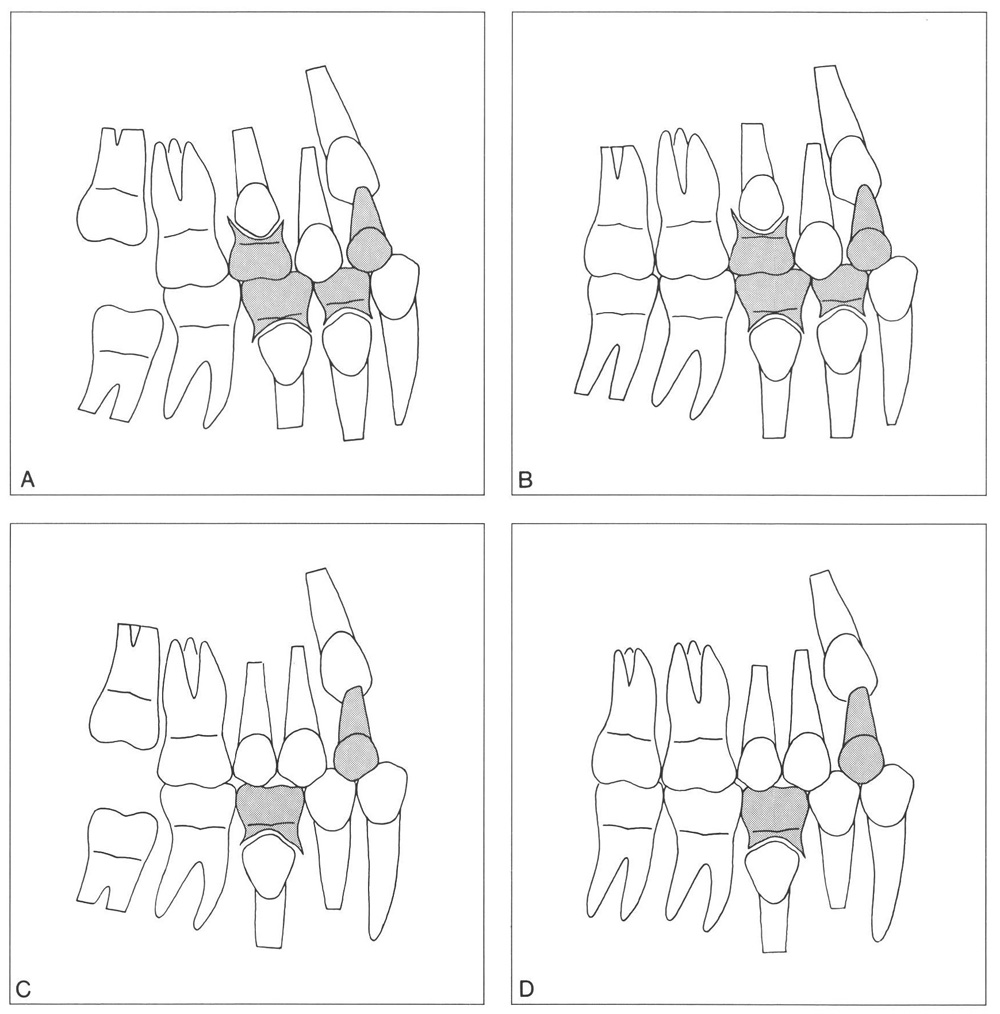
Fig. 3-5 Schematic presentation of some variations in the sequence of emergence of the posterior teeth, including the second permanent molars. (From Van der Linden.223)
A The mandibular permanent canine and the maxillary first premolar have emerged at approximately the same time. The second permanent molars in both jaws have not yet emerged.
B Emergence of the second permanent molars has taken place during or shortly after the transition of the mandibular canine and the emergence of the maxillary first premolar.
C The mandibular first premolar and permanent canine have emerged simultaneously. The same holds true for the two maxillary premolars. The second permanent molars have not yet emerged.
D The mandibular and maxillary second permanent molars have emerged before complete transition of the posterior teeth. The maxillary deciduous canine and mandibular second deciduous molar ha ve still to be replaced.
Deciduous molars are rarely resorbed prematurely in association with erupting adjacent permanent teeth. The crowns of the premolars are located more or less between the roots of their predecessors and seldom extend mesially or distally beyond the approximal crown surfaces of their predecessors. Shortage of available space for the eruption o/>
Stay updated, free dental videos. Join our Telegram channel
VIDEdental - Online dental courses


