3: Orofacial Pigmentation Disorders

Orofacial Pigmentation Disorders

Pigmented Lesions of Oral Mucosa

Oral mucosa can have a variety of discolorations like blue, black, brown or yellow because of endogenous pigments or exogenous materials. Recognition and diagnosis of pigmented lesions of the oral mucosa is an important aspect of clinical dental practice because it may be suggestive of an underlying systemic or metabolic disorder, or it can be an initial stage of malignancy. Therefore, dentists must determine the cause of pigmentation, ask relevant questions in the history to determine the cause and request appropriate investigations for early diagnosis and prompt treatment.

Molecular and Pathologic Correlation of Pigmentation

A pigment is any organic or inorganic coloring substance. A pigmented lesion can be defined as an area of altered coloration of the oral mucosa either because of physiologic or pathologic process because of deposition of endogenous or exogenous pigments or embedded foreign material in the tissues.

Exogenous substances cause oral mucosal pigmentation because these substances are embedded in the oral tissues either by direct trauma or iatrogenic implantation, amalgam being the most common. Cavity preparation of a tooth with an existing restoration (secondary caries) by a rotary bur can forcefully entrap some fine amalgam particles into the oral tissues. Amalgam can also enter oral tissues while removing old or fractured restorations. It can also accidentally enter into an extraction socket during extraction of a restored tooth. Amalgam particles can be left behind in the oral tissues after root canal treatment or retrograde amalgam filling. The silver particles from the embedded amalgam restorations slowly leach out and stain reticulum fibers causing gray/black discoloration of mucosa which is called as amalgam tattoo.

Lead pencils contain graphite which is another source of exogenous pigmentation which usually occurs in the palate. Rarely when the graphite fragment gets embedded in the palatal tissues following trauma, the mucosa covering this fractured fragment gets discolored and appears as a bluish black focal pigmented area.

In heavy metal poisoning also we can observe oral mucosal pigmentation because of formation of metal sulfides which get precipitated in inflamed areas of gingiva causing bluish black discoloration. Table 1 summarizes the type of exogenous substances that can cause oral mucosal pigmentation.

Table 1

Type of exogenous foreign materials causing pigmentation of oral mucosa

Source Color Etiology
Silver, amalgam Gray, black Iatrogenic implantation, trauma
Graphite Gray, black Tattoo, trauma
Lead, mercury, bismuth Gray Ingestion of paints, medicines, poisoning
Chromogenic bacteria Black, brown, green Superficial colonization

Oral mucosal pigmentation can also occur due to variety of endogenous pigments. Each of these endogenous pigments can result in a distinct oral discoloration suggestive of various disease processes which are described in Table 2. Blue, bluish red and purple type of oral pigmentation generally occur as a consequence of blood or vascular disorders. Black, brown or gray discoloration of the oral mucosa is because of melanin or hemosiderin pigments. Yellow discoloration of oral mucosa is because of bilirubin deposition or ingestion of large amount of beta-carotene. The pigmented lesions can either be localized or diffuse depending upon their etiology or disease process which they manifest.

Table 2

Type of endogenous pigments, the discoloration caused by them and disease process they indicate

Pigment Color Disease process
Hemoglobin Blue, red, purple Varix, hemangioma, Kaposi sarcoma, angiosarcoma, hereditary hemorrhagic telangiectasia
Hemosiderin Brown Ecchymosis, petechiae, thrombosed vein, hemorrhagic mucocele, hemochromatosis
Melanin Brown/black/gray Melanotic macule, nevus, melanoma, hormonal imbalance, drugs
Bilirubin Yellow Jaundice/liver disorder
Carotene Yellow Precursor of vitamin A

Blood pigments can cause red, bluish red or brown pigmentation of oral mucosa. These pigments are deposited into the connective tissues after lysis of erythrocytes causing extravasation of hemoglobin. The extravasation of blood into soft tissues is attributed to trauma, capillary fragility, platelet defects, or clotting disorders. This extravasated hemoglobin is acted upon by the enzymes to form hemosiderin, which is further broken down into bilirubin and biliverdin, all of which cause oral pigmentation. Therefore, the same endogenous pigments may result in different types of oral pigmentations over a period of time. An early hematoma is bluish red in color while a late hematoma becomes dark brown in color. Blood pigments are usually cleared from the skin or mucosa within 2 weeks.

Melanin causes blue or blue black discoloration of oral mucosa. Melanin is produced from an amino acid tyrosine by the melanocytes within the membrane-bound organelles called as melanosomes. These melanocytes are found in the basal layers of the oral epithelium. Melanin is also produced by the nevus cells, which originate from the neural crest cells and are found in the skin and mucosa. Color of pigmented lesions caused by melanin deposition may be brown, blue, gray or black, depending on the amount of melanin produced as well as on the location of melanin within the tissues.

Oral lesions can occur either due to excessive production of melanin pigment or due to proliferation of melanocytes. Sometimes there is no increase in the number of melanocytes but there is only an increase in the synthesis or production of melanin pigment. Over a period of time the capacity of cells to store melanin pigment is exceeded and then melanin spills into the underlying connective tissues into the adjacent basilar keratinocytes. This process is called as basilar melanosis and melanin incontinence.

In the oral cavity we can observe melanosis over the healed areas which had an earlier injury. When the traumatized oral epithelium regenerates, usually there is overproduction of melanin by the melanocytes that repopulate this region causing post inflammatory pigmentation.

Certain hormones and drugs can also influence production of melanin. The adrenal cortical-hypothalamic axis is affected by hypofunction of the adrenal cortex. As serum corticosteroid levels decreases, adrenocorticotropic hormone (ACTH) production by the posterior pituitary increases. ACTH also has a melanocyte stimulatory function so melanin production increases and it causes diffuse melanosis of the oral tissues. Therefore oral mucosal pigmentation can also occur in endocrine disorders. Certain drugs like minocycline can also increase the production of melanin within the cells causing diffuse pigmentation of oral mucosa and is called as drug-induced melanosis.

Oral pigmented lesions can also occur due to the proliferation of melanocytes as observed in the cases of nevi and melanoma. Melanocytic nevi develop during childhood and rarely arise in adult life. Most nevi originate from the melanocytes in the basal layer of epithelium and proliferate along the junction with the connective tissue. These are called as junctional nevi. Later these melanocytes drop off into the connective tissue to form islands of cells and are called as compound nevi. Later all these cells are completely detached from basal epithelium and form clusters in the dermis or submucosa and are called as intradermal nevi. Rarely do we observe blue nevi in oral cavity and they are comprised of spindle-shaped melanocytes which synthesize copious amounts of melanin pigment.

Occasionally, bluish discoloration of oral mucosa can be due to optical phenomenon as a consequence of accumulation of fluid within the epithelial layers. For example a ranula appears blue in color because the mucous absorbs most of the visible wavelength except blue.

In albinism the gene for tyrosinase is mutated. In vitiligo, depigmented patches are caused by diminished number of melanocytes. However, these entities are rarely seen in the oral cavity.

Metal Pigmentation

Pigmentation of the oral cavity caused by foreign materials or metals is called exogenous pigmentation. The importance of recognizing oral mucosal pigmentation caused by heavy metals lies primarily in the identification and treatment of the cause to avoid severe systemic toxic effects. Depending on the type of metal implicated, a number of systemic signs and symptoms may be associated with chronic exposure of metals.

Amalgam tattoo

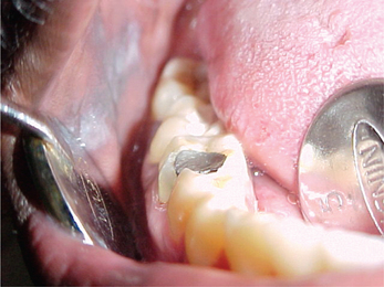
Amalgam tattoo is the most common solitary focal pigmentation lesion of the oral mucosa. These lesions are generally less than 1 cm (rarely amalgam tattoos can be large) and appear as flat, gray-black to blue-black color macules. They are usually found in close approximation to a restoration. Majority of the lesions are located on the buccal mucosa, gingiva and alveolar mucosa with mandibular region being more affected as compared to the maxillary region (Figure 1). All these lesions are asymptomatic and are discovered during routine dental examination. If particles are large enough they can be viewed with help of a radiograph (taken with reduced exposure parameters for soft tissues). The amalgam granules and fragments are found mainly in the lamina propria but were sometimes also seen in the submucosa. Histopathologically, we may observe a giant cell reaction surrounding these amalgam particles.

Mercury poisoning

Mercury poisoning can be because of acute or chronic exposure of mercury vapors. Clinical features include gastric disturbances, diarrhea, excitability, headache and mental depression. Patients complain of gastric disturbances, diarrhea, excitability, headaches and mental depression. Patient may have tremors of lips and extremities, dermatitis and nephritis.

Oral manifestations include increased salivation (ptyalism) as mercury is excreted in saliva. Tongue may be enlarged and painful (glossodynia). There may be hyperemia and swelling of gingiva, ulcerative stomatitis, loosening and exfoliation of teeth.

Acrodynia (Swift disease, Pink disease) is idiosyncratic reaction to large doses of amalgam. It is an uncommon mercurial toxicity reaction in which skin is also involved. It usually occurs after ingestion of mercury from powder, ammoniated mercury ointment, calomel lotion. It is mainly seen in the children below the age of 5–6 years. Manifestations are widespread involving the hand, feet, nose and cheeks which become red or pink in color, cold and clammy like a raw beef. Skin may have maculopapular rashes with pruritis. Patients may complain of irritability, photophobia and muscular weakness. Children may be able to remove their hair in patches.

Oral manifestations include profuse salivation, gingiva is painful and ulcerated, teeth may become loose and exfoliate and bruxism is a common finding.

Diffuse and Multiple Pigmented Lesions

Racial/physiologic pigmentation

Physiologic pigmentation of the oral tissues is clinically manifested as multifocal or diffuse melanin pigmentation with variable prevalence in different ethnic groups. Gingiva appears darker in color among Africans, Asians, Caucasians and Hispanics and other dark skinned people. The variability in the color of the oral tissues is not due to the difference in the number of melanocytes is same but this color difference is due to the difference in the activity of the melanocytes. In dark skinned people melanocytes are more active as compared to the fair skinned people.

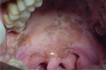
Physiologic pigmentation is seen as diffuse macular pigmentations that may be brown, gray or black in color and can appear anywhere in the mouth with buccal surface and gingiva being most commonly involved. On the gingiva it appears as well-demarcated, ribbon-like, dark brown continuous band that does not extend to involve the marginal gingiva. Occasionally pigmentation may also be seen on the tongue, lips and lingual gingiva as diffuse brown patches with ill-defined or diffuse borders (Figure 2).

Racial pigmentation mainly occurs in the childhood. However during early years it may not attract patient’s attention and may be observed after puberty. Racial pigmentation is asymptomatic and does not require any treatment.

Drug-induced pigmentation

Many medications when taken over a long period of time can cause oral mucosal pigmentation. Drug-induced pigmentation can be due to increased synthesis and accumulation of melanin pigments, deposition of the drug or its metabolites into the oral tissues or deposition of iron after damage to the dermal vessels (Table 3).

Table 3

List of drugs that can induce oral pigmentation

Bleomycin

Busulphan

Clofazimine

Chloroquine

Chlorpromazine

Cyclophosphamide

Doxorubicin

Estrogen

5-Fluorouracil

Gold

Hydroxychloroquine

Ketoconazole

Minocycline

Tetracycline

Quinacrine hydrochloride

Zidovudine

Chloroquine and other quinine derivatives which are usually used in the treatment of malaria and cardiac arrhythmia can cause pigmentation of oral tissues due to a direct stimulating effect on the melanocytes (Figure 3). According to some of the studies these drugs usually cause pigmentation of the palatal tissues.
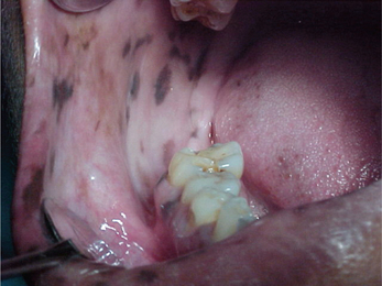
Minocycline is another drug causing pigmentation of oral tissues. It is a synthetic tetracycline that is commonly used in the treatment of acne vulgaris. Tetracycline causes pigmentation of only the bones and teeth but minocycline can also cause pigmentation of soft tissues. It usually causes diffuse brownish discoloration of the hard palate, gingiva, mucous membranes and tongue.

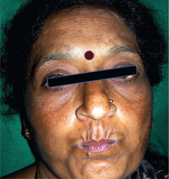
Oral pigmentation can also be due to intake of birth control pills. Chloasma is the term which is used to describe perioral and periorbital pigmentation in such patients (Figure 4). The pigmentation usually occurs as a diffuse brown macular pigmentation which is asymptomatic and lesions resolve upon cessation of drug intake. These lesions usually occur due to the hormonal changes which influence melanocyte stimulation.

Smoker’s melanosis

Tobacco smokers have more intense pigmentation of oral mucosa as compared to non-smokers. Smoker’s melanosis is more common in females. Women are more commonly affected than men because of synergistic effect between the female sex hormones and smoking. Smoking may cause oral pigmentation in light-skinned individuals and accentuate the pigmentation in dark-skinned patients. Clinically, there may be multiple diffuse brown pigmented macules of less than 1 cm in diameter. These lesions can occur anywhere in the mouth but may be usually localized on the attached labial anterior gingiva and the interdental papillae of the mandible. These lesions are completely asymptomatic and benign in nature (Figure 5).
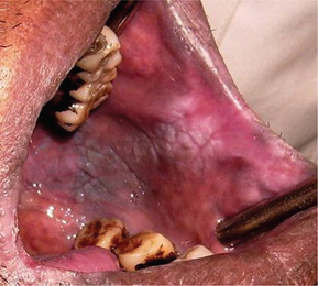
Microscopically there is evidence of basilar melanosis but there is no melanocyte proliferation. The increased melanin production may be a biologic defense mechanism against the noxious agents present in the tobacco smoke. The intensity of the oral pigmentation is directly related to the duration and amount of smoking. Smoker’s melanosis usually disappears within 3 years of smoking cessation. Biopsy must be advised whenever there is a surface elevation or increase in the pigment intensity or rapid increase in the size of the lesion.

Endocrine disorders

Adrenal cortical insufficiency can occur secondary to pathologic processes such as neoplasms that may cause damage to the adrenal glands. Due to the deficiency of the adrenocortical hormones in the blood there is an increased production of ACTH by the anterior pituitary gland. But ACTH stimulates the production of melanocyte-stimulating hormone which results in diffuse pigmentation of all the tissues. As a consequence the skin darkens and becomes bronzed. Also multifocal diffuse pigmentations appear in the mucous membranes of the oral cavity, conjunctiva, and genital regions.

In the oral cavity pigmentation may appear as diffuse brown patches on the gingiva, buccal mucosa, palate and tongue (Figure 6). This may resemble physiologic pigmentation. Physiologic pigmentation can be differentiated from pigmentation caused by Addison’s disease as the later develops and progresses during adult life and not present since birth. It is also accompanied by systemic manifestations like weakness, nausea and vomiting, abdominal pain, constipation or diarrhea, weight loss and hypotension. Oral pigmentation may be the first sign of Addison’s disease. Therefore complete examination must be done for patients with diffuse pigmentation of oral cavity and associated with systemic signs and symptoms. Patients presenting with such features should be sent for medical evaluation and laboratory tests to assess levels of ACTH, plasma cor-tisol and serum electrolytes.

A biopsy of these oral lesions shows acanthosis with silver-positive granules in the cells of the stratum germinativum. Melanin pigment can be seen in the basal layer of the epithelium. Management involves treatment of the underlying cause and corticosteroid replacement therapy.

A tumor of the posterior pituitary or certain small cell carcinomas can also secrete excessive amounts of ACTH which can cause pigmentation of oral tissues. In ACTH secreting tumors (paraneoplastic syndromes) the patient manifests features of the Cushing’s syndrome. In both these conditions there occurs diffuse pigmentation of oral tissues along with associated systemic features.

Café au lait pigmentation

It manifests as bronze or tan diffuse multifocal macular pigmentations that appear on the skin as well as the oral mucosa. These pale brown macules may vary considerably in size. They have widespread distribution and can occur on the face, neck or the oral cavity. Because of the pale brown color these lesions are called as Café au lait spots. It is usually associated with neurofibromatosis (von Recklinghausen’s syndrome), Albright’s syndrome (polyostotic fibrous dysplasia) and Peutz-Jeghers syndrome.

Café au lait pigmentations in neurofibromatosis have smooth borders and are associated with axillary freckling. It is an autosomal dominant inherited disease with multiple skin nodules.

In Peutz-Jeghers syndrome patients have intestinal polyposis along with oral macular pigmentations that appear around the mouth (Figure 7) and on the fingers. When such lesions are observed, a detailed history of the patient should be taken about gastrointestinal complaints as well as the family history for intestinal polyps. These pigmented melanotic spots do not require any treatment and are not associated with any risk for malignant transformation. However, the patient should be monitored for the development of internal m/>

Stay updated, free dental videos. Join our Telegram channel

Jan 12, 2015 | Posted by in Oral and Maxillofacial Radiology | Comments Off on 3: Orofacial Pigmentation Disorders

VIDEdental - Online dental courses

Get VIDEdental app for watching clinical videos