3 Noninfectious Papillary Lesions
Inflammatory Papillary Hyperplasia of the Palate
Clinical Findings
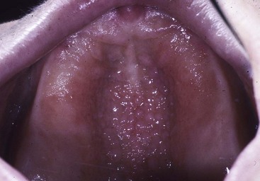
• Papillary hyperplasia of the palate usually occurs in adults wearing dentures; it is occasionally seen in patients with high-arched palates and mouth-breathing habit (with adenoidal facies); may be sensitive, especially if associated with erythematous candidiasis, because denture may act as a fomite; dryness from mouth-breathing also predisposes to candidiasis (Fig. 3-1).
• Pebbly excrescences appear on the palatal vault in a diffuse, symmetric distribution.
• Papillary lesions associated with human papillomavirus infection are discussed in Chapter 4.
Etiopathogenesis and Histopathologic Features
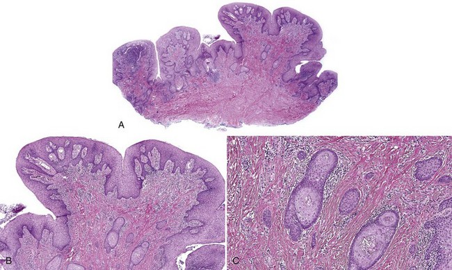
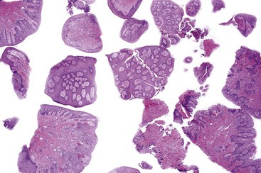
• There is proliferation of fibrous tissue in nodules with variable lymphoplasmacytic infiltrate; overlying epithelium may be hyperplastic and exhibit spongiosis and leukocyte exocytosis; pseudoepitheliomatous hyperplasia may be seen (Figs. 3-2 to 3-4).
• Presence of spongiotic pustules should always raise suspicion for candidiasis and a periodic acid–Schiff (PAS) stain with diastase should be performed.
Canger EM, Celenk P, Kayipmaz S. Denture-related hyperplasia: a clinical study of a Turkish population group. Braz Dent J. 2009;20:243-248.
Kaplan I, Vered M, Moskona D, et al. An immunohistochemical study of p53 and PCNA in inflammatory papillary hyperplasia of the palate: a dilemma of interpretation. Oral Dis. 1998;4:194-199.
Stay updated, free dental videos. Join our Telegram channel

VIDEdental - Online dental courses


