An Implant Is Not a Tooth
A Comparison of Periodontal Indices
Carl E. Misch
Dental implants are primarily used to replace teeth in a partial or complete edentulous patient or to retain removable prostheses. Therefore, the typical purpose of a dental implant is to act as an abutment for a prosthetic device, similar to a natural tooth root and crown. The restoring dentist usually designs and fabricates a prosthesis similar to one supported by teeth and, as such, also similarly evaluates and treats the dental implant as a natural tooth. Yet fundamental differences in the surrounding tissues and conditions between these devices are important to be recognized. For example, implants are different than natural teeth in that they do not decay, have no dental pulps to function as early indicators of disease or to cause lesions of endodontic origin, and have no periodontal membrane. The purpose of this chapter is to compare the periodontal indices for a natural tooth and an osteointegrated dental implant.
Literature Review
Several dental health criteria have been adapted for dental implants.1–12 A majority of reports that include clinical criteria to evaluate an implant include mobility; radiographic assessment of bone loss; and on occasion, gingival and plaque indices. Subjective criteria of discomfort and patient satisfaction also are mentioned.
The clinical criterion most commonly reported is the survival rate, or whether the implant is still physically in the mouth or has been removed.2 Proponents of this method say it provides the clearest presentation of the data; critics argue implants that should be removed because of pain, disease, or the inability to be restored still may be maintained yet wrongfully reported as successful. It should be noted that reports of traditional prostheses’ success supported by natural teeth follow a similar criterion: whether the restoration is still in the mouth. Therefore, survival rates rather than success rates are the most common method to report the “success” of dental implants or dental implant prostheses.
The American Dental Association Council on Dental Materials, Instruments, and Equipment states that consideration for an endosteal implant should be given to the evaluation of (1) durability; (2) bone loss; (3) gingival health; (4) pocket depth; (5) effect on adjacent teeth; (6) function; (7) esthetics; (8) presence of infection, discomfort, paresthesia, or anesthesia; (9) intrusion on the mandibular canal; and (10) the patient’s emotional and psychological attitude and satisfaction.3,4 One may reasonably state that factors controlled primarily by the dentist and the psychological attitude of the patient are not conditions influenced by the implant. As a result, these items should be considered separately and should not be computed in the evaluation of implant success.
Smith and Zarb suggested that patient comfort, sulcus depth, gingival status, damage to adjacent teeth, and violation of the maxillary sinus, mandibular canal, or floor of the nasal cavity are not attributable to the material or design of an implant.5 However, the sulcus depth and gingival status next to the implant indeed may be related to implant design or surface condition. For example, a smooth, polished collar placed below the bone contributes to crestal bone loss, which affects sulcus depth. The implant surface condition may allow bacteria to readily form on the surface of the implant body after crestal bone loss and may affect the gingival status of the implant. Hence, sulcus depth and gingival status are important indices for the evaluation of dental implants. Patient comfort, damage to adjacent teeth, and violation of anatomical structures are important to note but are not related to the design or material of an implant.
Periodontal indices are often used for evaluation of dental implants. A comparison of natural teeth and implants for each criterion provides insight into their differences in the health–disease continuum. After one understands the basis for evaluation, these criteria may then be used to establish a health–disease implant quality scale related to patient treatment.
This chapter addresses the more common periodontal indices for natural teeth and compares the following periodontal indices for dental implants: (1) longevity, (2) mobility versus rigid fixation, (3) percussion, (4) pain, (5) probing depths, (6) bleeding index, (7) crestal bone loss, (8) radiographic evaluation, (9) keratinized tissue, and (10) periimplant disease.
Longevity
Success criteria for endosteal implants have been proposed previously by several authors, including Schnitman and Shulman,6 Cranin et al.,7 McKinney et al.,8 Albrektsson et al.,9,11 and Albrektsson and Zarb.10 The success criteria by Albrektsson et al. was specific for implants with rigid fixation and is widely used today9 (Box 3-1). However, the Albrektsson et al. success criteria should be used primarily for implant studies or reports, not for individual implants. Assessing less than 0.2 mm vertical annual bone loss is difficult for each individual implant, and if 0.3 mm occurs in 1 year, it does not qualify as implant failure, in and of itself. In addition, the amount of crestal bone lost during the first year is not noted in this success criteria and may affect the sulcus depth and environment for the longevity of the implant.
Minimum survival rates for a success criteria suggested in the Albrektsson et al.9 guideline are low compared with present-day reports and do not consider the prosthesis survival related to implant longevity. This report states the minimum implant success rate is 85% for 5 years and 80% for 10 years. However, the proposed criteria do not evaluate the prosthesis. The consideration of a minimum implant survival rate should be in the context of the final prosthesis survival. For example, many early reports indicated that a fixed prosthesis in a completely edentulous arch may be supported by four implants. In a study of 25 patients with 25 prostheses supported by only four implants, there would be 100 implants. A 75% implant success rate could result in 0% prosthesis success if each patient lost only one implant. An 85% implant 5-year survival rate with this treatment plan still would cause almost half the implant restorations to fail. Of course, this survival rate is not acceptable. Implant survival by itself is not an acceptable criterion to evaluate an implant system, and studies should also include the implant restoration.
Implant “success” is not as relevant for clinical implant evaluation as the quality of health. “Success” is not used for the evaluation of a tooth. Instead, a quality of health is more relevant for the clinical criteria. The clinical criteria for optimum to satisfactory health for implants established by Misch and accepted by the International Congress of Oral Implantologists Pisa Consensus Conference evaluates both implant and prosthesis survival and suggests a minimum of 90% prosthesis survival for 10 years1,13 (Table 3-1). Of course, these rates require even greater implant survival, and overengineering the support system often is required to obtain this goal. For example, if eight rather than four implants support a full arch fixed prosthesis, possibly one or two implants may be lost, and the same prosthesis still may be used without additional implants and only slight modification of the restoration. For example, if 25 patients were each restored with eight implants to support a full-arch restoration and if each patient lost one implant, the survival rate of the prosthesis still may be 100% and the implant survival rate 87%.
TABLE 3-1
Implant Quality
| Scale Group | Management | Clinical Conditions |
| I. Success (optimum health) | Normal maintenance | No pain or tenderness upon function 0 mobility <2 mm radiographic bone loss from initial surgery Probing depth <5 mm No exudate history |
| II. Survival (satisfactory health) | Reduction of stresses Shorter intervals between hygiene appointments Gingivoplasty Yearly radiographs |
No pain 0 mobility 2–4 mm radiographic bone loss Probing depth 5–7 mm No exudate history |
| III. Survival (compromised health) | Reduction of stresses Drug therapy (antibiotics, chlorhexidine) Surgical reentry and revision Change in prosthesis or implants |
No pain upon function 0 mobility Radiographic bone loss >4 mm Probing depth >7 mm May have exudate history |
| IV. Failure (clinical or absolute failure) | Removal of implant | Pain upon function Mobility Radiographic bone loss >  length of implant length of implantUncontrolled exudate No longer in mouth |
Suggested Criteria for Implant Success1
• Implant quality scale* of 1, 2, or 3 with a survival rate better than 90% at 10 years.
From International Congress of Oral Implantologists, Consensus Conference, Pisa, Italy, 2008.
Computed data of dental implant survival or success should include all implants inserted, not just the implants restored or those successfully loaded after 1 year. An implant placed and left submerged should most often be included in initial or surgical implant failure.
The time of implant failure is also relevant. For the patient and doctors involved in treatment, a 10% implant failure rate before fabrication of the prosthesis is far better than to have 5% implant failure rate after delivery of the restoration.14 The most common time for an implant failure is within the first 18 months after loading. Hence, most often the final restoration has been delivered and in function.
Mobility
Natural Tooth versus Implant Support Systems
Compared with an implant, the support system of a natural tooth is better designed to reduce the biomechanical forces distributed to the tooth/restoration and the crestal bone region. The periodontal membrane, biomechanical design of the tooth root and material, nerve and blood vessel complex, occlusal material (enamel) and surrounding type of bone blend to decrease the risk of occlusal overload to the natural tooth system.15
Tooth Movement
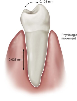
The tooth exhibits normal physiologic movements in vertical, horizontal, and rotational directions. The amount of movement of a natural tooth is related to its surface area and root design. Therefore, the number and length of the roots; their diameter, shape, and position; and the health of the periodontal ligament (PDL) primarily influence a tooth’s mobility. A healthy tooth exhibits zero clinical mobility in a vertical direction. Actual initial vertical tooth movement is about 28 microns and is the same for anterior and posterior teeth.16 The vertical movement of a rigid implant has been measured as 2 to 3 microns under a 10-lb force and is due mostly to the viscoelastic properties of the underlying bone.17
Muhlemann found that horizontal tooth movement may be divided into initial mobility and secondary movement.18 The initial mobility is observed with a light force, occurs immediately, and is a consequence of the PDL. Initial horizontal tooth mobility is greater than initial vertical movement. A very light force (500 g) horizontally moves the tooth. The initial horizontal mobility of a healthy, “nonmobile” posterior tooth is less than that of an anterior tooth and ranges from 56 to 75 microns, which is two to nine times the vertical movement of the tooth. Initial horizontal mobility is even greater in anterior teeth and ranges from 70 to 108 microns in health16,19 (Figure 3-1).
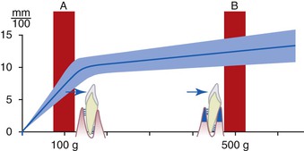
The secondary tooth movement described by Muhlemann occurs after the initial movement when greater forces are applied. When an additional force is applied to the tooth, a secondary movement is also observed, which is related directly to the amount of force. The secondary tooth movement is related to the viscoelasticity of the bone and measures as much as 40 microns under considerably greater force18(Figure 3-2).
Implant Movement
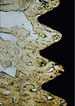
Rigid fixation indicates the absence of clinical mobility of an implant tested with vertical or horizontal forces less than 500 g. Rigid fixation is a clinical term. Osseointegration is a histologic term defined as bone in direct contact with an implant surface at the magnification of a light microscope20 (Figure 3-3). Over the years, these two terms have been used interchangeably, and implant abutment support is most predictable with rigid fixation. Lack of implant mobility (IM) does not always coincide with a direct bone–implant interface.8 However, when observed clinically, rigid fixation usually means that at least a portion of the implant is in direct contact with bone, although the percentage of bone contact cannot be specified.21 A mobile implant indicates the presence of connective tissue between the implant and bone.
Lack of clinically observable movement does not mean the true absence of any movement. For example, a “nonmobile” posterior natural tooth actually moves horizontally 56 to 73 microns. The human eye does not perceive this movement. The anterior teeth, which often have slight clinically observable movement, actually move approximately 0.1 mm. A healthy implant moves less than 73 microns; thus, it appears as zero clinical mobility (rigid fixation).
Just as a natural tooth, the implant–bone interface exhibits more lateral than apical movement. Sekine et al. evaluated the movement of endosteal implants with rigid fixation and found a range of 12 to 66 microns of movement in the labiolingual direction.17 Komiyama reported 40 to 115 microns of implant movement in the mesiodistal direction under a force of 2000 g (≈4.5 psi) and a labiolingual range of 11 to 66 microns.22 The greater implant movement in the mesiodistal dimension corresponds to the lack of cortical bone between the implants in this direction compared with the thicker lateral cortical plates present in the labiolingual dimension. Rangert et al. suggested that part of this implant movement may also be due to component flexure of the implant abutment and screw.23 The mobility of implants varies in direct proportion to the load applied and the bone density and reflects the elastic deformation of bone tissue.
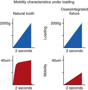
Sekine et al. applied a gradually increasing load over a 2-second period to a tooth and an implant. The teeth moved immediately with a light load (primary tooth movement) and less with an additional load (secondary tooth movement). The implant did not move when the tooth had its primary tooth movement. A heavier force caused the implant to gradually move, similar to the secondary tooth movement17 (Figure 3-4). These mobility characteristics corroborate the findings of Fenton et al., who applied a 500-g load for 4 seconds to maxillary anterior teeth and osseointegrated implants.24 Whereas the implants were displaced a mean of 10 microns with a rapid elastic return (less than 1 millisecond), the teeth showed a mean displacement of 57 microns with a prolonged viscoelastic return.
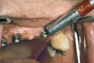
Increased tooth mobility may be caused by occlusal trauma or bone loss. Increased tooth mobility alone is not a criterion of periodontal health or pathology. Unlike a tooth, for which mobility is not a primary factor for longevity, mobility is a primary determining factor for implant health.20 Rigid fixation is also an excellent indicator of the implant health status because it is an easy, objective test. As such, rigid fixation is usually the first clinical criterion evaluated for a dental implant. The techniques to assess rigid fixation are similar to those used for natural tooth mobility. Two rigid instruments apply a labiolingual force of approximately 500 g, and no observed mobility indicates rigid fixation25 (Figure 3-5).
The amplitude of tooth mobility may be rated from 0 to 4, where 0 is normal mobility from physiologic movement; 1 is detectable increased mobility; 2 is visible mobility up to 0.5 mm; 3 is severe mobility up to 1 mm; and 4 is extreme mobility, including vertical movement.25 This same gradient may be used for oral implants with slight modification. As Box 3-2 depicts, IM-0 corresponds to the absence of clinical mobility, IM-1 demonstrates detectable increased movement, IM-2 is visible mobility movement up to 0.5 mm, IM-3 is severe horizontal mobility greater than 0.5 mm, and IM-4 is visible horizontal and vertical movement. The IM scale was used frequently for plate (blade) form implants or disc implants because a clinical goal was for slight mobility when joining the device to natural teeth. However, the goal for root form implants always should be rigid fixation and IM-0 status.
A natural tooth with primary occlusal trauma exhibits an increase in clinical mobility and radiographic PDL space. After the cause of trauma is eliminated, the tooth may return to zero clinical mobility and a normal radiographic appearance. This scenario is not predictable around an implant. The dentist should not restore an implant with any clinical mobility, because the risk of failure is great. However, after the prosthesis is completed and IM-1 develops, the risk is small to evaluate the implant for a few months and decrease almost all stress during this time frame. Implants with slight detectable mobility of approximately 0.1 mm of horizontal movement (IM-1), similar to the mobility of a healthy central incisor, on occasion may return to rigid fixation and zero mobility. However, to reachieve rigid fixation, the implant should be taken completely out of occlusion for several months. Chances improve to return rigid fixation to an implant if no mobility is noted before the implant is placed into function.
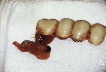
An implant with horizontal movement greater than 0.5 mm (IM-3) is at much greater risk than a tooth. A root form implant with greater than 0.5 mm horizontal mobility (IM-3) or any vertical mobility (IM-4) should be removed to avoid continued bone loss and future compromise of the implant site (Figure 3-6).
On occasion, an implant that was rigid may spin in the bone at stage II uncovery, when the implant abutment is threaded into position.26 The weak bone–implant interface is broken when the shear forces of adding an abutment and screw are placed on the implant body. If this occurs, the implant cover screw should be reinserted and the implant allowed to “reintegrate” with the bone. The chances are greater than 75% that 3 additional months of healing will allow the implant to reestablish a bone–implant interface if it was present before the abutment was added.27 At the reinsertion of the abutment, a lesser torque is used initially, and a counter torque technique is used (so the rotational force on the abutment screw is not converted to the bone–implant interface, so the interface does not strip again). After an additional time of progressive loading, the abutment screw may be tightened as usual, although a counter torque method on the abutment is still suggested.
The Periotest (Gulden-Medizinteknik, Bensheim an der Bergstrasse, Germany) is a computer-mechanical device, developed by Schulte, that measures the dampening effect or attenuation degree against objects by developing a force of 12 to 18 N against a pistonlike device, which then measures the distance the piston recoils into the chamber after striking an object.28 A soft surface or mobile object gives higher recordings than a hard or rigid object. The recordings range from negative 8 to positive 50 numbers.
Teeth with zero clinical mobility have typical Periotest ranges from 5 to 9. The degree or absence of clinical movement around an implant corresponds to values ranging from −8 to +9, or a range of 17 units (Figure 3-7). The bone density around the implant may be correlated with Periotest numbers. Whereas softer bone types give higher numbers, harder bone around the implant results in lower numbers. A nondestructive resonance frequency analysis technique to measure implant stability and osteointegration has also been introduced to the profession and provides similar valuable information as to the clinical movement and bone density around implants.29,30 These devices greatly aid the dentist’s tactile senses.
The Periotest device has also been used as a clinical tool to evaluate slight changes in implant rigid fixation or to identify prostheses that become partially unretained.31–33 Because the restoration does not need to be removed to evaluate the implant, this device may more easily be used to evaluate an implant long term.
Percussion
Percussion often is used on teeth to determine which tooth is sensitive to function or is beginning to abscess. In the past, percussion was used to evaluate the presence of rigid fixation for osteointegrated implants.20 However, percussion is an indicator neither of clinical health nor of rigid fixation for osteointegrated implants. The ringing sound that occurs on percussion only corresponds to the presence of some bone at the interface, because 2 mm of bone and 16 mm of bone–implant interface sound almost identical. Percussion may be used to diagnose pain or tenderness with an implant but is misleading if used to determine the status of rigid fixation.
Pain
Subjective findings of pain, tenderness, and sensitivity are common dental conditions that the dentist treats as part of a general practice. Pain and tenderness are subjective criteria and depend on the patient’s interpretation of the degree of discomfort. Pain is defined as an unpleasant sensation ranging from mild discomfort to excruciating agony. Tenderness is more an unpleasant awareness of the region. A natural tooth often becomes hyperemic and sensitive to cold as the first indicator of a problem. A tooth with a more serious condition becomes sensitive to heat and painful to percussion, indicating pulpitis. Dental emergencies usually are associated with pain, and the dentist is adept at its diagnosis and treatment planning.
An implant rarely is troubled by the subjective criteria of pain or sensitivity after initial healing. The implant does not become hyperemic and is not temperature sensitive, and the early warning signs and symptoms of a traumatic occlusal problem may not be present. This criterion is less contributory to implant health determination.
After the implant has achieved primary healing, absence of pain under vertical or horizontal forces is a primary subjective criterion. Usually (but not always), pain does not occur unless the implant is mobile and surrounded by inflamed tissue or has rigid fixation but impinges on a nerve. The most common condition that causes discomfort from an implant is when a loose implant abutment is entrapping some of the soft tissue in the abutment–implant connection. After the soft tissue in the region is eliminated and the abutment is secured, the discomfort subsides. When the abutment–implant connection is secure and pain is present, consideration is given to an implant body fracture.
On rare occasions, an implant may cause discomfort during function, although a clinical examination is unable to identify a cause. The persistent presence of pain during percussion or function on properly inserted implants and components often requires removal of the implant even in the absence of mobility. Because pain is a subjective criterion, the dentist asks the patient to relate the pain from the implant site on a scale of 1 to 10, with 1 being a slight aggravation and 10 being the most intense pain the patient can perceive. When the patient reports a pain level greater than 5, the dentist should strongly consider removal of the implant.
Whereas pain from rigidly fixated implants is rare and is observed as an early problem, pain from a mobile implant may occur early or late in treatment. In either case, the condition rarely improves. Pain on loading of rigid implants has been observed more often on immediately loaded implants compared with those healing unloaded for an extended period.
Implant sensitivity or mild tenderness rather than pain in a rigid implant is also most unusual and signals a more significant complication for an implant than for a tooth. Tenderness during function or percussion usually implies healing in the proximity of a nerve or, on rare occasions, bone stress beyond physiologic limits.
If the implant tenderness immediately after surgery and loading (immediate loading) occurs when the implant is in the proximity of the mandibular canal, the implant may be unthreaded 1 mm and reevaluated for a decrease in symptoms after 3 or more weeks. If the tenderness of a rigid fixated implant is after stage I healing and is not due to surgical encroachment on an anatomical landmark, attention is first brought to the soft tissue and prosthetic components. If this is not the cause, treatment then consists of the elimination of as much stress on the implant or prosthesis as is possible for 3 or more weeks. The dentist especially should address occlusion and parafunctional habits in the presence of implant sensitivity. Most often the prosthesis should be modified to reduce a cantilever of occlusal contacts. On occasion, additional implants may be placed and the restoration remade to dissipate the forces. The tenderness may be decreased with these procedures but rarely is eliminated. Instead, the dentist notifies the patient of the poor prognosis and asks whether the tenderness is significant enough to warrant the removal of the implant. It should be emphasized this condition is rare and has been observed only a few times by the author in more than 30 years.
On occasion, an implant body may fracture from fatigue. Fatigue is related to the amount of force, the number of cycles, the strength of the material, the diameter of the component, and the number of implants splinted together. This condition is similar to a fractured root. In any case, radiographic evidence of the fracture may be difficult to ascertain. Percussion and forces up to 500 g (1.2 psi) with a bite stick are used clinically to evaluate a tooth or implant for pain or discomfort. Percussion and heavy biting on a wood stick associated with pain are clinical indices. In these cases, the implant is most often removed.
Probing Depths
Probing depths around teeth are an excellent proven means to assess the past and present health of natural teeth. The increasing sulcus depth around natural teeth is related to disease and bone loss.25 However, probing depth indices used to evaluate dental implants are more controversial because relating implant sulcus depth to health is not always directly related.
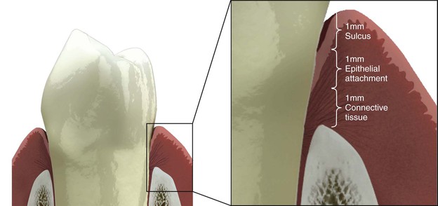
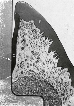
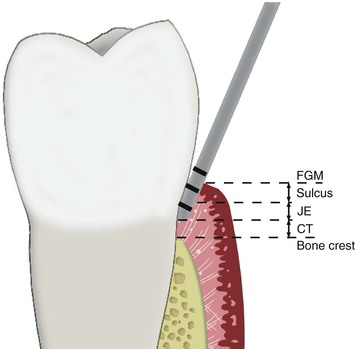
For a natural tooth, the surrounding soft tissue has an average biological width of 2.04 mm between the depth of the sulcus and the crest of the alveolar bone.34 It should be noted the biological “width” is actually a height dimension with a greater range in the posterior region compared with the anterior and may be greater than 4 mm in height.35 In teeth, it is composed of a connective tissue attachment (1.07 mm average) above the bone and a junctional epithelial attachment (JEA) (0.97 mm average) at the sulcus base, with the most consistent value among individuals being the connective tissue attachment (Figure 3-8).
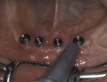
The sulcular regions around an implant and around a tooth are similar in many respects. The rete peg formation within the attached gingiva and the histologic lining of the gingiva within the sulcus are similar in implants and teeth.36 A free gingival margin forms around a tooth or implant with nonkeratinized sulcular epithelium, and the epithelial cells at its base are also similar to teeth and implants, with junctional epithelial cells for both (Figure 3-9). However, a fundamental difference characterizes the base of the gingival complex around teeth. Whereas a tooth has two primary regions that make up the biological width, an implant only has one.
When probing next to a tooth, the probe not only measures the sulcus depth but also penetrates and measures the JEA.37 The junctional epithelial “attachment” of a tooth is not a true attachment. A periodontal probe easily separates the hemidesmosomal close approximation of the epithelial cells. High-volume air from a syringe may blow it off, plaque destroys it, and the placement of impression string in the sulcus displaces it. In other words, the mucopolysaccharide close approximation of the hemidesmosome found in the JEA is not an attachment (Figure 3-10).
The connective tissue attachment zone of the “biological width” around a tooth prevents the probe from penetrating deeper into the sulcus and allows gingival fibers of the connective tissue attachment zone to establish direct connection with the cementum of the natural tooth. It acts as a physical barrier to the bacteria in the sulcus to the underlining periodontal tissues. Eleven different gingival fiber groups comprise the connective tissue attachment zone observed around a natural tooth and tissue: dentogingival (coronal, horizontal, and apical), alveologingival, intercapillary, transgingival, circular, semicircular, dentoperiosteal, transseptal, periosteogingival, intercircular, and intergingival.25 At least six of these gingival fiber groups insert into the cementum of the natural tooth: the dentogingival (coronal, horizontal, and apical), dentoperiosteal, transseptal, circular, semicircular, and transgingival fibers. In addition, some crestal fibers from the periodontal fiber bundles also insert into the cementum above the alveolar bone. These Sharpey fibers form a true attachment to the tooth. They prevent a periodontal probe from invading the PDL space and delay the ingress of plaque.
James and Schultz were first to begin a systematic study to investigate the biological seal phenomenon of the soft tissue around dental implants.36 Hemidesmosomes from the JEA region help form a basal lamina–like structure on the implant, which can act as a biological seal.38 However, collagenous components of the linear body cannot physiologically adhere to or become embedded into the implant body.39 The hemidesmosomal seal has a circumferential band of gingival tissue to provide mechanical protection against tearing.40 However, the mucopolysaccharide layer is less adherent to an impl/>
Stay updated, free dental videos. Join our Telegram channel
VIDEdental - Online dental courses


