(b) 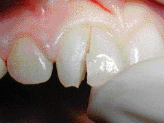
All the upper incisor and canine teeth responded positively to thermal and electric pulp test. No other cracks or fractures were found. A slight haematoma was visible on the inner aspect of the upper lip adjacent to the 11. Pocket depths and tooth mobility were within normal limits. There was no evidence of intrusion or lateral luxation of any of the teeth.
Two radiographs at different vertical angulations were taken of upper incisor teeth. A crown fracture was detected in the 11 (Figures 3.5.2a–b). The fracture line extended from the distal third of the incisal edge mesially and apically to the marginal bone level on the mesial aspect of the tooth.
Figure 3.5.2 (a) The first periapical radiograph demonstrates a fracture line extending from the distal third of the incisal edge of the crown to the mesial marginal bone level. The two stronger (darker) lines in the radiograph represent the labial and palatal extents of the fracture. No horizontal root fracture can be detected, (b) the second periapical radiograph taken from another projection verifies the findings of the first radiograph.
(Courtesy of Haapasalo et al. Visual Endodontics 2010 multimedia.)
(a) 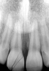 (b)
(b) 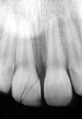
Why in trauma cases are multiple radiographs taken?
At least two radiographs are necessary as otherwise there is a chance that the fracture line will remain undetected. In situations where the diagnosis still remains uncertain, additional radiographs may be required. In this case, two obvious and one weak fracture line could be detected on the radiographs. This is not an indication of multiple fractures but it reflects the path of one fracture line. As the apical extent of the fracture line extended just onto the root surface, the case could be defined as crown fracture.
The root development of all the anterior teeth was complete. This would be expected based on the age of the patient. The radiograph indicated that the fracture line involved the pulp. This is common in young patients as the pulp horns extend relatively close to the incisal edge.
Why did moving/touching the fractured segment cause sharp pain?
Teeth with a complicated fracture (as in this case) are usually very sensitive to even the slightest touch. Exposure of the pulp means that if the fractured piece is touched and moved, direct mechanical pressure is applied to the nerve receptors in the pulp; this causes a strong, sharp sensation of pain. Some of the pain will also arise from gingival fibres attached to the fragment.
Can the tooth be retained?
Although the fracture line extended below the marginal gingiva, the prospects of treating and saving the tooth were still favourable. This is important as the age of the patient was a contraindication for implant treatment.
Diagnosis and treatment planning
A provisional diagnosis of complicated crown fracture was reached. The initial treatment plan was the removal of the fractured part of the crown and assessment of the residual tooth. Verification of the diagnosis included confirmation of a pulpal exposure and whether the root was involved. If assessment was favourable, emergency endodontic treatment and temporary restoration of the crown would follow.
What are the treatment options in this case?
The possible endodontic treatments for the tooth are summarized in Table 3.5.1. There was a direct choice between pulp preservation or extirpation. Due to the fracture extending onto the root surface, there was concern about being able to provide a good seal to eliminate leakage from the margin. There were further concerns regarding future restoration if the pulp was conserved. If the root development had not been complete, pulpotomy would have been the first choice of treatment in order to allow further development of the root and strengthening of the tooth. However, the root was fully formed and pulpectomy was considered a better option.
Stay updated, free dental videos. Join our Telegram channel
VIDEdental - Online dental courses


