(b) 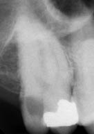
What did the radiograph (Figure 3.3.1b) reveal about the 17?
- Mesio-occlusal restoration.
- Extensive distal decay.
- No evidence of a periapical radiolucency.
- Good marginal bone levels.
Diagnosis and treatment planning
The diagnosis was reversible pulpitis associated with a carious tooth.
It is prudent to investigate this tooth by removal of the existing restoration under local anaesthetic to assess the extent of the caries. If the tooth is restorable and the pulp is not exposed, the tooth may be restored with a direct plastic restoration such as composite or amalgam. Further treatment will have to be carried out prior to restoration if caries excavation results in a pulpal exposure.
What were the potential treatment options for the patient if the pulp is exposed?
- Leave alone.
- Pulp capping or partial pulpotomy.
- Root canal treatment.
- Extraction.
Direct pulp capping, partial pulpotomy and pulpectomy are the options available when managing the exposed vital (healthy) pulp. Direct pulp capping is a procedure in which the pulp is covered with a protective dressing or base placed directly over the site of the exposure. The only difference between pulpotomy and pulp capping is that with the former, additional tissue is removed from the exposure site. A ‘full pulpotomy’ involves removal of the entire coronal pulp to the level of the root canal entrance, while with a ‘partial pulpotomy’ only the superficial layer of damaged and/or inflamed tissue is removed, prior to placement of the base.
When is it possible to conserve the pulp?
In theory, a reversibly inflamed pulp exposed by either trauma or caries may be conserved. However, the problem is associated with being able to assess accurately the degree of pulpal inflammation, as this will influence treatment outcome. For traumatic exposures, vital pulp treatment is the treatment of choice, particularly, if root formation is incomplete. However, carious exposures are generally treated by pulpectomy (i.e. root canal treatment) as the pulp is presumed to be extensively inflamed due to a sustained bacterial infection. A marked difference in treatment outcome has been noted between inflamed and non-inflamed pulps after vital pulp treatment.
Options other than pulpectomy may be considered-as in this case-where the tooth was asymptomatic, responded positively to electrical/thermal sensibility testing and there was no radiographic evidence of a periapical radiolucency. These clinical signs indicated that the pulp was healthy, and in such cases, pulp capping or partial pulpotomy may be an option.
What are the advantages of pulp (preservation) capping?
Pulp capping is less time-consuming, less costly and less invasive than root canal treatment (which would be the only alternative if the patient wanted to retain his tooth). In addition, the patient has avoided a crown.
From a biological perspective, pulpal preservation treatment maintains the defensive properties of the pulp such as secondary and tertiary dentine deposition, and the pulp-dentine proprioception is unaffected, which should reduce the likelihood of overloading the tooth and subsequent fracture.
What factors are required for successful vital pulp capping?
- Healthy or reversibly inflamed pulp.
- Careful technique ’ attention to haemorrhage control and placement of capping material.
- Prevention of microleakage ’ capping material and permanent restoration must have good sealing properties to prevent further bacterial contamination.
The patient did not wish to lose the tooth. It was not possible to determine the actual extent of the decay prior to investigation. The patient was informed of this uncertainty, the likelihood of pulpal exposure, and the treatment options should this occur. The decision was made to investigate the 17 initially and, if exposed, consider a pulp capping or pulpotomy procedure.
If we proceed with a pulp capping/pulpotomy, what should we advise the patient?
- Discuss options and explain the rationale for treatment.
- That the technique is conservative, but that further treatment (i.e. root canal treatment) may be necessary in the future should the tooth become symptomatic.
- Regular reviews will be required.
- Some initial cold sensitivity can be expected.
Treatment
The tooth was isolated with rubber dam, and a caulking material was used to provide further resistance to the ingress of saliva (Figure 3.3.2a). The existing mesio-occlusal amalgam restoration was removed, and the decay was removed from the periphery of the cavity with a round bur in a slow handpiece. Finally, the decay was removed from the pulpal aspect of the cavity with an excavator. The tooth was examined and found to be restorable, but removal of the decay resulted in pulpal exposure.
Stay updated, free dental videos. Join our Telegram channel
VIDEdental - Online dental courses


