Radiological differential diagnosis – describing a lesion
Introduction
As a result, many of these pathological conditions resemble one another closely. This often creates considerable confusion. Fortunately, the sites where the lesions develop, how they grow and the effects they have on adjacent structures tend to follow recognizable patterns. As mentioned in Chapter 19, it is the recognition of these particular patterns that provides the key to interpretation and the formation of a radiological differential diagnosis.
Detailed description of a lesion
Site or anatomical position
This should be stated precisely, for example the lesion(s) could be:
• Localized to the mandible, affecting:
• Localized to the maxilla, affecting:
• Originating from a point or epicentre relative to surrounding structures, e.g.:
In the mandible, so-called odontogenic lesions develop above the inferior dental canal, while non-odontogenic lesions develop above, within or below the canal. Thus some conditions have a predilection for certain areas whilst others develop in one site only. For example, radicular dental cysts develop at the apices of non-vital teeth, while so-called fissural bone cysts develop only in the midline. The site or anatomical position of a lesion may therefore provide the initial clue as to its identity.
Size
Conventionally, the lesion is sized in one of two ways including:
• Measuring the dimensions in centimetres
• Describing the boundaries, i.e. ‘the lesion extends from … to … in one dimension and extends from … to … in the other dimension’, as shown in Fig. 25.1.
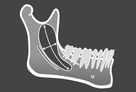
 up to the sigmoid notch, and from the anterior border of the ramus down to the ID canal,’ or ‘It is approximately 6 cm × 2 cm’.
Stay updated, free dental videos. Join our Telegram channel
VIDEdental - Online dental courses


