24: Venipuncture Technique

chapter 24 Venipuncture Technique

The preparation of the equipment for a continuous intravenous (IV) infusion and venipuncture technique is described in this chapter.

PREPARATION OF EQUIPMENT
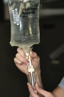
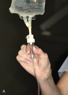
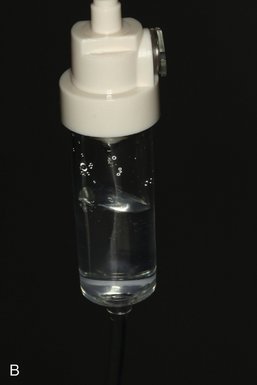
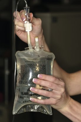
1. The armamentarium discussed in Chapter 22 is laid out and removed from its packaging.
3. If a winged infusion set is used, it is removed from its box and attached to the end of the IV tubing (Figure 24-2). The protective sheath covering the needle is left on. If an indwelling catheter is used, it is not attached to the IV tubing.
8. The drip chamber on the IV tubing should be filled approximately halfway with IV fluid. If it is not, the chamber can be squeezed and released to draw additional fluid into it (Figure 24-5). If the chamber is overfilled to the extent that it is impossible to visualize individual drops of solution as they exit the IV bag, the bag and drip chamber are inverted and the drip chamber squeezed to force fluid back into the IV bag (Figure 24-6). Care should be taken to prevent the entry of air bubbles into the IV tubing at this time.

Additional equipment required for venipuncture includes the following:

PREPARATION FOR VENIPUNCTURE

The patient is asked to visit the restroom, if necessary, before the start of the venipuncture. Once the IV has been started and central nervous system (CNS)–depressant drugs administered, it will be more difficult for the patient, even assisted by office staff, to accomplish a visit to the restroom.

The patient is seated comfortably in the dental chair. A semireclined to supine position is recommended as physiologically superior to the upright position for maintenance of cerebral blood flow and adequate respiration.

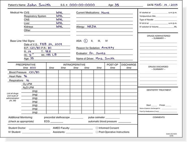
Preoperative vital signs are recorded on the patient’s sedation record sheet (Figure 24-7). Included should be the blood pressure, heart rate and rhythm, respiratory rate, and O2 saturation. The patient’s baseline vital signs have been recorded at a prior visit to the dental office.

The blood pressure cuff (sphygmomanometer) is placed on the left arm and left in place throughout the IV procedure. The right arm should be used if the dentist is left-handed.

The patient’s arms (without the tourniquet in place) are scanned for obvious veins. Often veins will be made readily visible if the arm is permitted to hang down below the level of the patient’s heart for a few minutes because this augments venous distention (Figure 24-8). One of the goals of the preoperative visit was to determine whether the patient has suitable veins for the IV procedure.

On occasion, a patient who had very visible, superficial veins at the preoperative visit will appear in the office on treatment day with no obvious veins. This is explained by the presence of a greater degree of anxiety with attendant higher levels of circulating catecholamines (producing peripheral vasoconstriction). When veins are not readily apparent, the patient should be asked at what site on the arm he has previously had blood drawn successfully. Sometimes a patient will boast that “they had to try four times before they found a vein” or that “three people had to try before they succeeded.” This should alert the dentist that a difficult venipuncture might be in the offing. However, such a statement by the patient may also be used to the dentist’s advantage. If, by using “special care,” the dentist is able to successfully complete the venipuncture on the first or second attempt, the patient will be more confident in that dentist’s overall ability. Several methods of distending veins are available and are discussed later.

If the Trieger test for evaluation of recovery is to be used (see Chapter 5), the baseline Trieger test is now completed by the patient. Any other monitoring devices, such as the pulse oximeter, capnograph, pretracheal stethoscope, or electrocardioscope, are now placed on the patient.

Because of the possibility of accidental inoculation of health professionals with viral and other organisms found in some patients’ blood, universal precautions are essential in situations involving potential contact with blood. All persons working with the venipuncture should wear masks, glasses, and gloves. Gloves must be worn throughout the procedure, from the point of preparation until the IV is removed, the bleeding stops, and a bandage is in place.

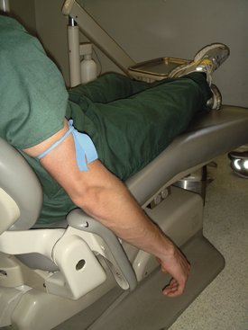
A tourniquet is placed on the limb (arm or leg) selected for venipuncture. On the arm, the tourniquet is placed superior to the antecubital fossa. The commonly used soft rubber tubing (e.g., Penrose drain) is applied in a slipknot (Figure 24-9). The tourniquet should be sufficiently tight to prevent venous drainage from the arm without obstructing arterial flow into the arm. A radial pulse should still be palpable with the tourniquet in place.

When a blood pressure cuff is used as a tourniquet, the pressure in the cuff is raised and maintained at a point between the patient’s systolic and diastolic pressures (e.g., ∼120 mm Hg if the blood pressure is 140/90 mm Hg). This produces venous distention in the same manner as the tourniquet.

The patient is then asked to repeatedly open and close his or her hand into a fist. Muscular activity forces more blood into the veins, allowing additional arterial blood to enter into the limb, further distending the veins. Once the veins have been distended, the patient is asked to keep the fist clenched until venipuncture has been successfully completed.

At this point, most persons will have one or more readily visible veins; however, some others may not have visible or even palpable veins. Several methods are available to increase venous distention.

image

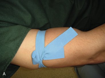
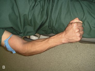
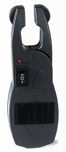
Figure 24-10 Vein locating device (www.med1online.com).

(Courtesy Venoscope, L.L.C., Lafayette, LA.)

When N2O-O2 is used as an aid during venipuncture, the patient should be sedated as described in Chapter 15 (titrated) and returned to a nonsedated state before the administration of any IV drugs.

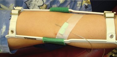
Once the vein chosen for cannulation has been adequately distended, the site must be prepared. Physical restraints are seldom required in adults because most patients rarely object strenuously to venipuncture (although they may not “like it”). When a vein in the antecubital fossa or wrist is selected, an elbow or wrist immobilizer should be placed before the start of the venipuncture if a rigid metal needle is to be used (Figure 24-11). An immobilizer is unnecessary if a flexible catheter is to be inserted.

Some experts recommend that any solution used to cleanse the injection site be warmed to body temperature. Because the arm is warm, it is more sensitive to cold so that the/>

Stay updated, free dental videos. Join our Telegram channel

Jan 5, 2015 | Posted by in General Dentistry | Comments Off on 24: Venipuncture Technique

VIDEdental - Online dental courses

Get VIDEdental app for watching clinical videos