20
Introduction to Cone Beam Computed Tomography
This technology, first described in 1998 for applications in dentistry (Mozzo et al., 1998), employs a cone-shaped X-ray beam emanating from a point source coupled with a planar digital sensor. During image acquisition, both the radiation source and sensor rotate around the patient, who is stationary. There are two classes of cone beam systems currently, ones that employ small fields of view with dimensions of less than 8 cm, and large fields of view with dimensions of greater than 8 cm upward to 30 cm.
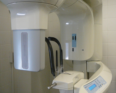
Unlike intraoral digital imaging, the anatomy of the area imaged is recreated in three dimensions rather than two. The three-dimensional (3D) elements that recreate the anatomy are referred to as cube-shaped volume elements or voxels. Small field of view systems (Figure 20.1) employ pixel dimensions as low as 0.076 mm while the larger field machines employ pixel dimensions of between 0.20 mm and 0.40 mm.
Figure 20.1 Small field of view CBCT system (Kodak 9000 3D, Carestream, Rochester, New York).

For endodontic applications (Nurbakhsh et al., 2011; Wang et al., 2011), which often require imaging resolutions at the level of the tooth and/or their supporting structures, the smaller field, higher resolution machines may be of greater value.
Prior to image acquisition, the area of interest is centered within the imaging volume. The center of the field determines what tissues may potentially be irradiated, and this has a bearing on patient radiation doses. Although radiation dose considerations are of particular concern in 3D imaging, they are less for small volume cone beam computed tomography (CBCT) than large volume CBCT or />
Stay updated, free dental videos. Join our Telegram channel

VIDEdental - Online dental courses


