2 Examination of mouth, jaws, temporomandibular region and salivary glands
Figure 2.1a Portable miniature operative light.
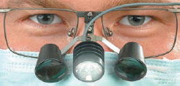
Figure 2.1b ENT headlight.
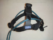
Figure 2.2a Teeth and gingivae.
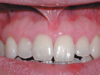
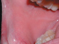
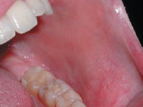
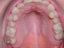
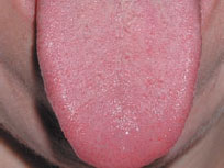
Figure 2.2f Tongue ventrum and floor of mouth.
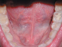
Figure 2.3 Common diseases.
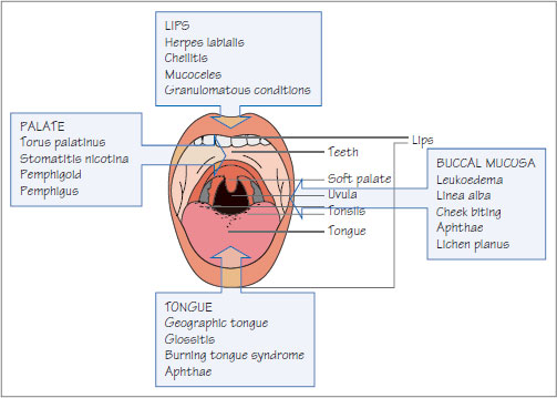
Figure 2.4 Toluidine blue.
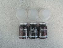
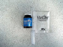
Figure 2.5 Chemiluminescent illumination system (ViziLite).
The lips are best first inspected. Complete visualization intraorally requires a good light; this can be a conventional dental unit light, or special loupes or ENT light (Figures 2.1a and b). If the patient wears a dental appliance, this should be removed to examine beneath.
Mouth
The dentition and occlusion should be examined. Study models on a semi- or fully-adjustable articulator may be needed. This is discussed in basic dental textbooks.
All mucosae should be examined, beginning away from the focus of complaint or location of known lesions. Labial, buccal, floor of the mouth, ventrum of tongue, dorsal surface of tongue, hard and soft palate mucosae, gingivae and teeth should be examined in sequence, recording lesions on a diagram (Figures 2.2a–f). Lesions are described as in Table 2.1.
Some conditions are found only in, or typically in, certain sites (Figure 2.3).
Mucosal lesions are not always readily visualized and, among attempts to aid this, are:
- toluidine blue (vital) staining
- chemiluminescent illumination
- fluorescence spectroscopy and imaging
Toluidine blue staining (Figure 2.4) stains mainly pathological areas blue. The patient rinses for 20 seconds with/>
Stay updated, free dental videos. Join our Telegram channel

VIDEdental - Online dental courses


