2: Assessing patients

Assessing patients

Overview

This chapter describes the basic principles of assessing a dental patient. A history should include significant medical and social facts as well as the dental problem. An initial extraoral examination covers both the visual appearance of the patient and features such as swellings and nerve dysfunction. Once these aspects are completed, the intraoral examination will attempt to identify any lumps or swellings and to differentiate these into dental and non-dental origins. Features such as ulcers and motor or sensory nerve dysfunction will also be noted before the detailed examination of the troublesome tooth or teeth. The physical examination of the teeth is described. Specific investigations must be chosen for their suitability, both in terms of the usefulness of the results and the medicolegal aspects of their use. For example, both HIV testing and the use of X-rays have implications beyond the results that they provide. The relative merits of the various investigations are described.

2.1 History

A full and accurate history is of paramount importance in assessment of a patient. In some cases, the history may provide the diagnosis, while in the remainder, it will give essential clues to the nature of the problem. The approach to history taking needs to be tailored to the type of complaint being investigated.

It is important to have a systematic approach to taking a history. A consistent series of questions will avoid inadvertently missing an important clue. Use ‘open’ rather than ‘closed’ (those usually eliciting a yes/no response) questions wherever possible to avoid leading the patient. Record the patient’s own responses rather than paraphrasing. The history will cover:

Medical history

‘Now some questions about your general health.’ This is obviously important. Some medical conditions may have oral manifestations, while others will affect the manner in which dental treatment is delivered. Even if the patient volunteers that they are ‘fit and healthy’ when you say you are going to ask them a few medical questions, you must persist and enquire specifically about key systems of the body:

See Chapter 3 for a more detailed discussion of the medical aspects of dental care.

2.2 Extraoral examination

Like history taking, examination necessitates a systematic approach. As a general rule, use your eyes first, then your hands to examine a patient. Start with the extraoral examination before proceeding to examine the oral cavity.

Take time to look at the patient. This may seem obvious but will identify swellings, skin lesions and facial palsies. Facial pallor may indicate anaemia, or that the patient may be about to faint. This process of observation will start while you are taking the history. Visual areas would cover:

Palpation would cover:

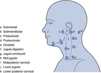
Lymph node examination

The major lymph nodes of the maxillofacial region and neck are shown in Fig. 2.1. The submental, submandibular and the internal jugular nodes (jugulo-digastric and jugulo-omohyoid node being the largest) are of particular importance because these receive lymph drainage from the oral cavity. Examination of the nodes should be systematic, although the order of examination is not critically important. To palpate the nodes, the examiner should stand behind the patient while he/she is seated in an upright position. Use both hands (left hand for the left side of the patient, etc.). A common sequence would be to start in the submental region, working back to the submandibular nodes then further back to the jugulo-digastric node (see Fig. 2.1). Then continue by palpation of the parotid region downwards to the retromandibular area and down the cervical chain of nodes. When a node is perceived as enlarged, record the texture: a hard node of a metastasising malignancy contrasts well with a tender, softer node in an inflammatory process.

Problem-specific examination

The examination will be made in the light of the symptoms reported by the patient, but the examiner may detect swelling, sensory or motor disturbance that the patient has not noticed.

Swelling/lump

The procedure for examination of a swelling or a lump must encompass a range of observations, including:

Consistency can be informative, ranging from the soft swelling of a lipoma, through ‘cartilage hard’ pleomorphic adenomas and ‘rubbery hard’ nodes in Hodgkin’s disease, to the ‘rock hard’ nodes of metastatic malignancy. Tenderness and warmth on palpation usually indicates an inflammatory process, while neoplasms are commonly painless unless secondarily infected. Fluctuation indicates the presence of fluid. To assess fluctuation, place two fingers on the swelling and press down with one finger. If fluid is present, the other finger will record an upward pressure. Pulsation in a swelling will indicate direct (i.e. it is a vascular lesion) or indirect involvement (i.e. in immediate contact) of an artery.

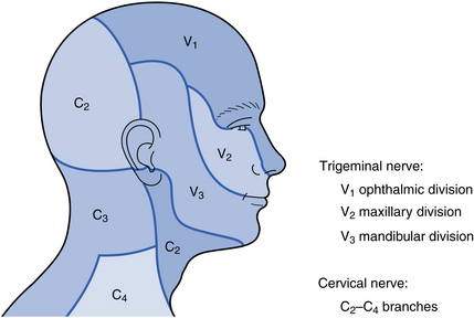
Paraesthesia/anaesthesia

The presence of sensory disturbance is usually identified initially by the patient in the history. It is important to identify the extent of the affected area and the degree of alteration in sensation. It is best to use a fairly fine, but blunt-ended, instrument for this at first, for example, the handle of a dental mirror. First, run the instrument gently over what is assumed to be a normal area of skin so that the patient knows what to expect. Then repeat this over the symptomatic area, asking the patient to say whether they can feel anything. Record the area of altered sensation in the notes using a drawing.

The degree of alteration in sensation can be assessed by using different ‘probes’. A teased-out piece of cotton wool can be used or, where anaesthesia appears to be profound, a sharp probe can be (carefully) tried.

The extent of the area of paraesthesia or anaesthesia will tell you the particular nerve, or branch of a nerve, involved (Fig. 2.2). This will, in turn, inform you about the possible location of the underlying lesion. For example, a patient with disturbed sensation of the upper lip has a lesion affecting the maxillary division of the trigeminal nerve. If this is the sole site of sensory deficit, it suggests a lesion closer to the terminal branches of this cranial nerve (e.g. in the maxillary sinus). In contrast, if sensory deficiencies are simultaneously present in other branches of the nerve, it suggests that the lesion is more centrally located.

Paralysis/motor disturbance

While paralysis or motor disturbance may be reported as a symptom by the patient, it may initially be identified during an examination. In the maxillofacial region, the motor nerves that are likely to be under consideration are the facial nerve, the hypoglossal nerve and the nerves controlling the muscles that move the eyes.

Disturbance in function of the facial nerve will result in effects on the muscles of facial expression. Paralysis of the lower face indicates an upper motor neurone lesion (stroke, cerebral tumour or trauma). Paralysis of all the facial muscles (on the affected side) indicates a lower motor neurone lesion. The latter is seen in a large number of conditions but, for the dentist, important causes include Bell’s palsy (Fig. 2.3), parotid tumours, a misplaced inferior dental local anaesthetic and trauma.
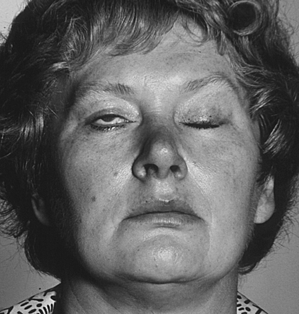
2.3 Intraoral examination

Again, a systematic approach is essential to avoid being distracted by the first unusual finding you encounter. The examination must include lips, cheeks, parotid gland orifices, buccal gingivae, lingual gingivae and alveolar ridges in edentulous areas, hard palate, soft palate, dorsal surface of the tongue, ventral surface of the tongue, floor of mouth, submandibular gland orifices and, finally, the teeth. Different clinicians will have their own sequence of examination, but it is the thoroughness of the examination that is important, not the order in which the regions of the mouth are examined.

Once the general intraoral examination is complete, a problem-specific examination can proceed. This is tailored to the clinical problem.

Stay updated, free dental videos. Join our Telegram channel

Jan 9, 2015 | Posted by in Oral and Maxillofacial Pathology | Comments Off on 2: Assessing patients

VIDEdental - Online dental courses

Get VIDEdental app for watching clinical videos