Introduction to radiological interpretation
• To identify the presence or absence of disease
• To provide information on the nature and extent of the disease
To achieve these objectives and maximize the diagnostic yield, interpretation should be carried out under specified conditions, following ordered, systematic guidelines.
Essential requirements for interpretation
The essential requirements for interpreting dental radiographs can be summarized as follows:
• Understanding the nature and limitations of the black, white and grey radiographic image
• Knowledge of what the radiographs used in dentistry should look like, so a critical assessment of individual image quality can be made
• Detailed knowledge of the range of radiographic appearances of normal anatomical structures
• Detailed knowledge of the radiographic appearances of the pathological conditions affecting the head and neck
• A systematic approach to viewing the entire radiograph and to viewing and describing specific lesions
Optimum viewing conditions
For film-captured images these include:
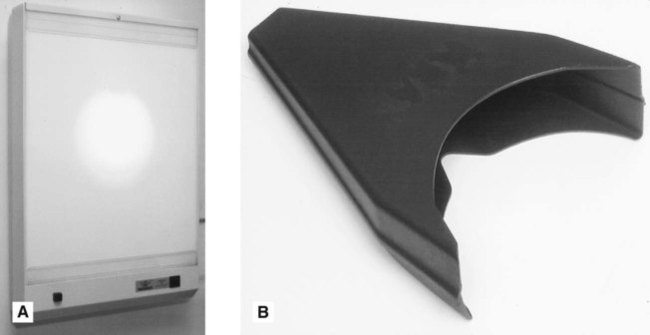
• An even, uniform, bright light viewing screen (preferably of variable intensity to allow viewing of films of different densities) (see Fig. 19.1)
• A quiet, darkened viewing room
• The area around the radiograph should be masked by a dark surround so that light passes only through the film
• Use of a magnifying glass to allow fine detail to be seen more clearly on intraoral films
These ideal viewing conditions give the observer the best chance of perceiving all the detail contained within the radiographic image. With many simultaneous external stimuli, such as extraneous light and inadequate viewing conditions, the amount of information obtained from the radiograph is reduced. Film-captured radiographs should be viewed once they have dried as films still wet from processing may show some distortion of the image.
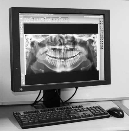
Digital images should be viewed on bright, high-resolution monitors in subdued lighting (see Fig. 19.2). Table 19.1 outlines the minimum and ideal specifications of the monitor (image display device) as recommended in the UK by the Health Protection Agency and by the Royal College of Radiologists.
Table 19.1
Summary of the minimum and ideal specifications for the monitor when viewing digital images
| Minimum specification | Ideal specification | |
| Screen resolution | ≥1280 × 1024 (~1.3 megapixels) | ≥1500 × 2000 |
| Screen size (viewable diagonal) | ≥42cm (~17″) | ≥50cm (~20″) |
| Maximum luminance | >170 cd/m2 | ≥500cd/m2 |
| Luminance contrast ratio | ≥250 : 1 | ≥500 : 1 |
| Greyscale bit depth | 8-bit greyscale (24-bit colour) | ≥10-bit greyscale |
The nature and limitations of different radiographic images
The importance of understanding the nature of different types of radiographic images – film-captured or digital (depending on the type of image receptor used) and their specific limitations was explained in Chapter 1. How the visual images are created by processing – chemical or computer – was explained in Chapter 5. Revision of both of these chapters is recommended. To reiterate, the final image whether captured on film or digitally is ‘a/>
Stay updated, free dental videos. Join our Telegram channel
VIDEdental - Online dental courses


