EYE AND ORBIT
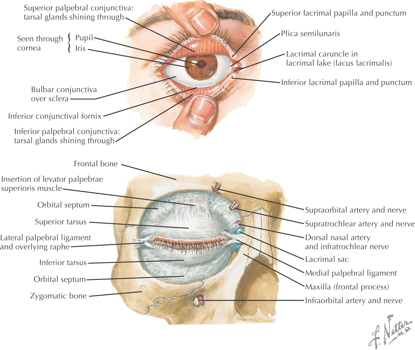
Overview and Topographic Anatomy of the Orbit
Overview and Topographic Anatomy of the Orbit
GENERAL INFORMATION
Orbit: a pyramid-shaped bony recess in the anterior part of the skull, lined by periosteum called the periorbital fascia
Contents include:
• Eye—organ associated with vision
• Ophthalmic division of the trigeminal nerve
• Ophthalmic artery and branches
• Superior and inferior ophthalmic veins
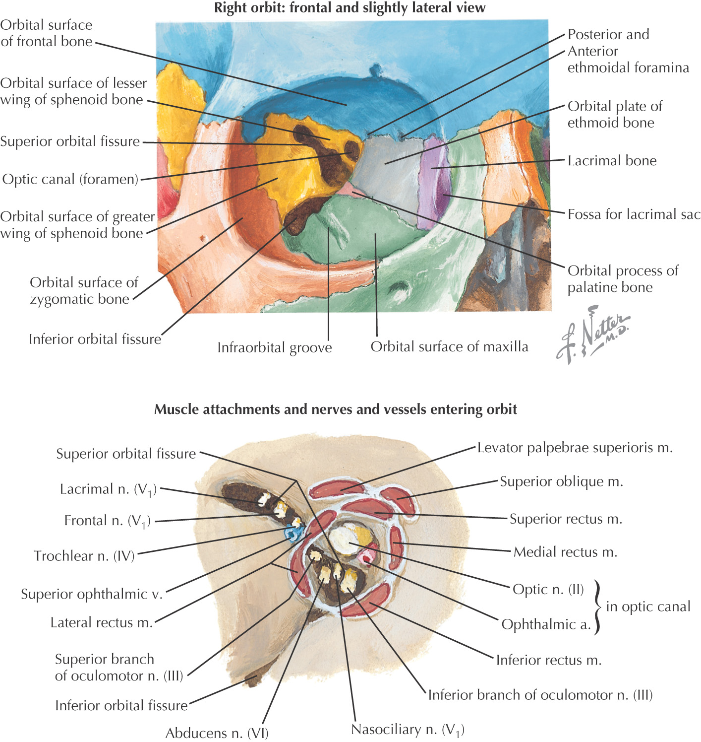
Osteology of the Orbit
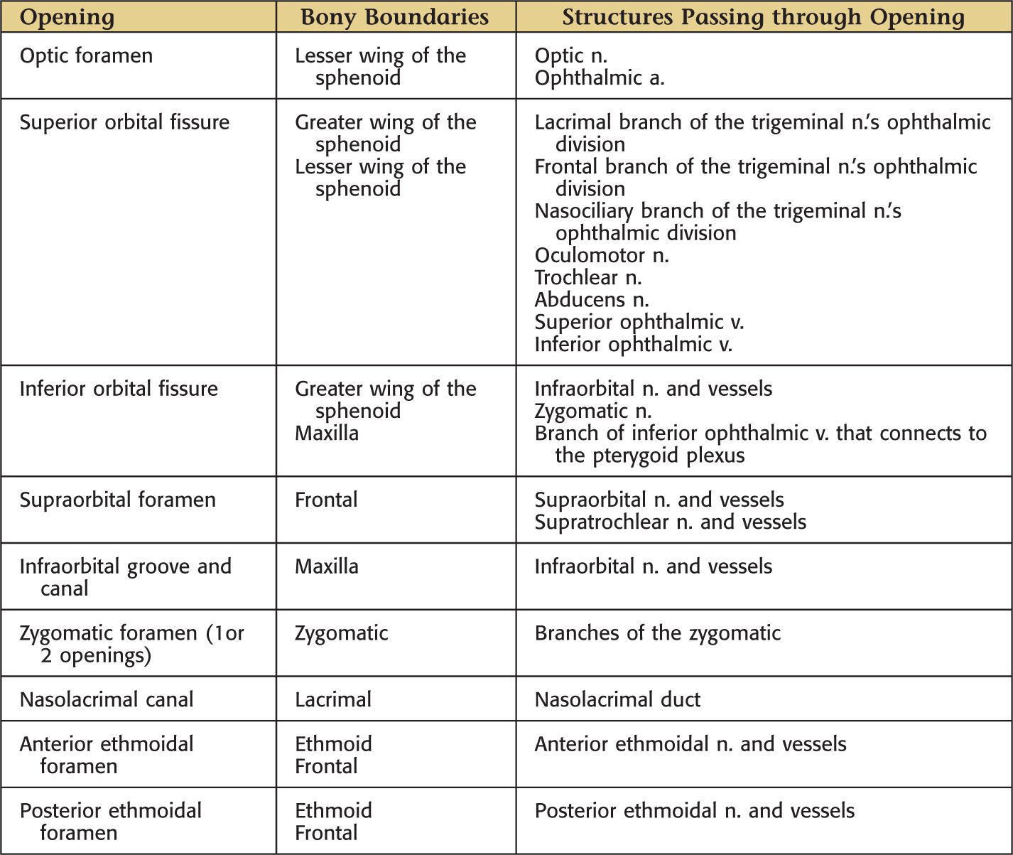
OPENINGS IN THE ORBIT
BONES CREATING THE ORBITAL MARGIN
WALLS OF THE ORBIT
|
Superior |
|
|
Inferior |
|
|
Medial |
|
|
Lateral |
Contents of the Orbit
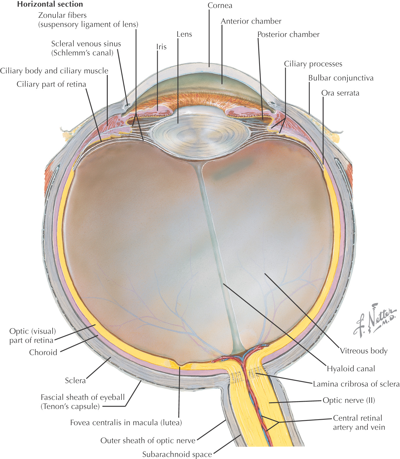
EYE
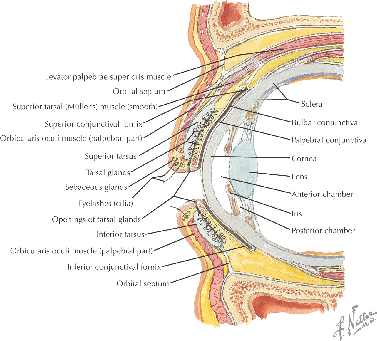
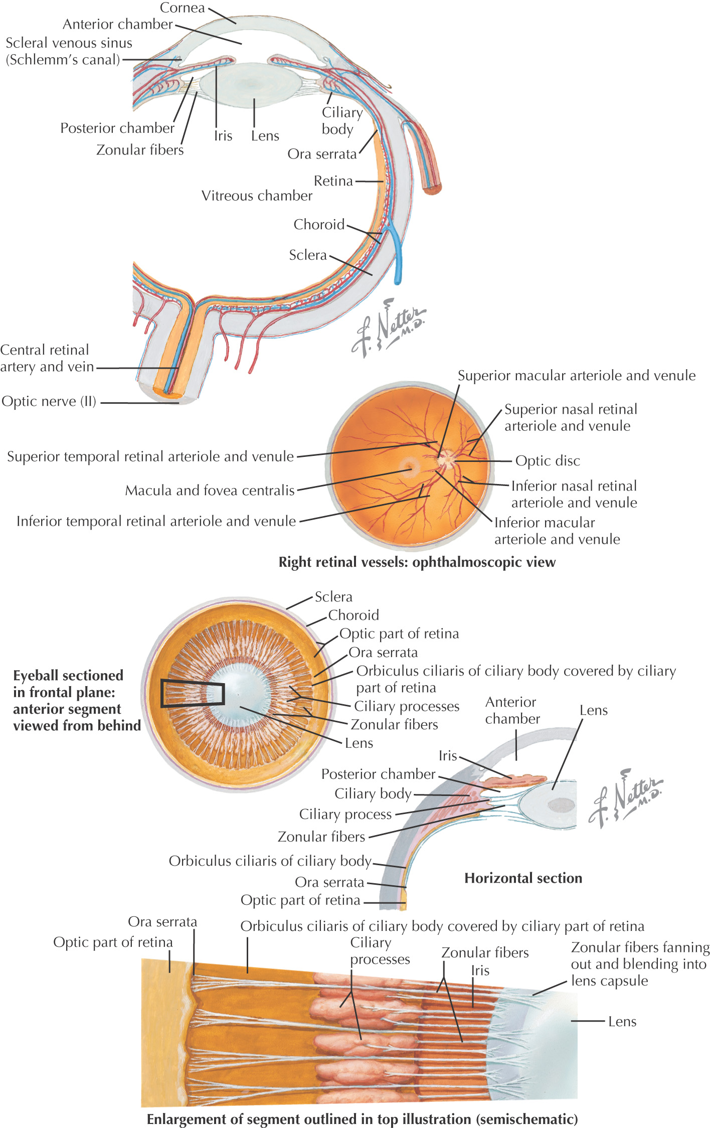
Eye: a spherical globe with a diameter of approximately 2.5 cm that lies in the orbit’s anterior portion
Surrounded by a thin capsule called the fascia bulbi (Tenon’s capsule):
Composed of 3 coats:
• Sclera
• Retina
Divided into an anterior and a posterior segment:
• Separated into anterior and posterior chambers by the iris
• Intraocular pressure is measured in the anterior segment, normally 10 to 20 mm Hg
Stay updated, free dental videos. Join our Telegram channel

VIDEdental - Online dental courses


