18
The Dentogingival Apparatus
Periodontal health is essential for successful prosthodontic treatment outcomes. This chapter describes basic periodontal aspects requiring analysis before embarking on restorative treatment.
Superficial Anatomy
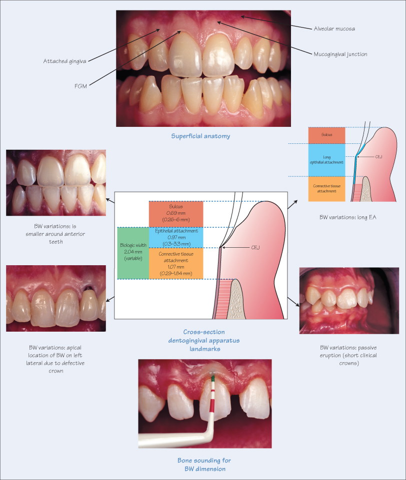
Viewed from the facial or buccal aspect, the soft tissue envelope around the teeth is distinguished into the gingiva and alveolar mucosa. Starting from the tooth junction, the gingiva is further divided into the free gingival margin (FGM) and attached gingiva, terminating apically at the mucogingival junction with the alveolar mucosa. The attached gingiva is keratinised, tenacious and resilient to withstand masticatory function, while the alveolar mucosa is unattached and non-keratinised.
The dimensions and texture of the attached gingiva vary enormously, not only between individuals, but also around specific teeth. The width (apico-coronal dimension) varies from 0.5 mm to 8 mm, and the average thickness (bucco-lingual dimension) is 1.4 mm. The function of the attached gingiva is to protect the soft tissues during oral function, and its absence, especially around implants, has been shown to increase the propensity to inflammation.
Dentogingival Apparatus
In cross-section, the dentogingival apparatus can be divided into three components: epithelium, connective tissue and bone. In a healthy periodontium, the sulcus starts at the FGM and ends at the epithelial attachment (EA). The depth of the sulcus (or gingival crevice) varies from 0.3 mm to 6 mm depending on numerous factors such as location on the tooth (deeper sulcus interproximally). The EA extends apically from the sulcus to the cemento-enamel junction (CEJ), ranging from 0.3 mm to 3.3 mm. />
Stay updated, free dental videos. Join our Telegram channel
VIDEdental - Online dental courses


