Cone beam computed tomography (CBCT)
Main indications
The developing dentition
• Localization of an unerupted tooth (small FOV)
• Assessment of external resorption in relation to unerupted teeth (small FOV)
• Localized assessment of an impacted tooth (small FOV)
• Assessment of cleft palate (small or medium FOV)
• Planning complex orthodontic/surgical management of maxillofacial skeletal abnormalities (medium or large FOV).
Restoring the dentition
If conventional imaging proves inadequate, CBCT may be indicated for:
• Assessment of periodontal infra-bony defects and furcation lesions (small FOV)
• Periapical assessment (small FOV)
• Assessment of root canal anatomy in multi-rooted teeth (small FOV)
• Planning surgical endodontic procedures (small FOV)
• Endodontic treatment complicated by resorption lesions, combined perio-endo lesions, perforations and atypical pulp anatomy (small FOV)
• Assessment of dental trauma (suspected root fracture) (small FOV).
Surgical applications
• Assessment of lower third molars where an intimate relationship with the inferior dental canal is suspected (small FOV)
• Assessment of unerupted teeth (small FOV)
• Cross-sectional imaging prior to implant placement (small or medium FOV)
• Assessment of pathological lesions affecting the jaws (small or medium FOV) including cysts, tumours, giant cell lesions and osseous dysplasias
• Assessment of facial fractures where soft tissue detail is not required (medium or large FOV)
• Planning orthognathic surgery to obtain three-dimensional datasets of the craniofacial skeleton (medium or large FOV)
• Assessment of the bony elements of the TMJ (small or medium FOV)
Clinical examples illustrating many of these indications are included in later chapters.
Equipment and theory
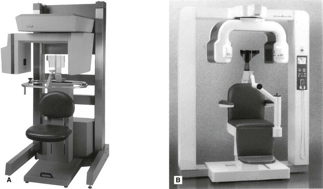
Multiple CBCT machines are currently available with new, upgraded models launched regularly by most manufacturers of X-ray equipment. Almost all modern machines resemble panoramic units, as shown in Fig. 16.1.
All equipment employs a cone-shaped X-ray beam (rather than the flat fan-shaped beam used in conventional CT as described in Chapter 18) and a special detector (e.g. an image intensifier linked to a charge-coupled device (CCD) or, more commonly, an amorphous silicon flat panel). The scanning/image creation process divides into three stages:
Stage 1 – Data acquisition
The patient is positioned with the unit (as described later). The equipment orbits around the patient in a 180°, 270° or 360° rotation, taking approximately 5–40 seconds, and in one cycle or scan, images a cylindrical or spherical volume referred to as the field of view (FOV). As all the information/data is obtained in the single scan, the patient must remain stationary throughout the exposure. As described earlier, the FOV can vary in size enabling small, medium or large volumes of a patient to be imaged. Using a large field of view (e.g. 15 cm3) most of the maxillofacial skeleton fits within the cylindrical or spherical shape and is imaged in the one scan as shown in Fig. 16.2.
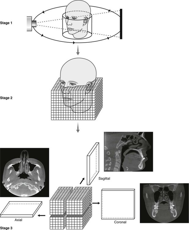
Stage 2 – Primary reconstruction
Having obtained data from the one scan, the computer then divides the volume into tiny cubes or voxels (ranging in size between 0.076 mm3 and 0.4 mm3) and calculates the X-ray absorption in each voxel. As with pixels in two-dimensional digital imaging, described in Chapter 5, each voxel is allocated a number and then allocated a colour from the grey scale from black through to white. Typically one scan contains over 100 million voxels. The overall image resolution clinically of hard tissues (teeth and bones) is generally very good in CBCT imaging, although measured spatial resolution (3–4 line pairs/mm) is not yet as good as two-dimensional film-based or digital imaging (10–25 lp/mm) (see Chapter 4). Using a smaller voxel size potentially increases the spatial resolution but increases the radiation dose. Even so CBCT cannot be used to examine the soft tissues in detail because of the size of the kV and types of detectors used and the amount of scatter. Essentially all that can be seen is the outline of the soft tissues where it interfaces with air.
Stay updated, free dental videos. Join our Telegram channel
VIDEdental - Online dental courses


