15: Traumatology

Traumatology

In order to treat an injured patient, it is important to know the effects of trauma on the hard and soft tissues and to plan proper emergency treatment, which will be followed by definitive treatment and maintenance.

Epidemiology

The teeth most frequently affected by traumatic events are the upper central incisors (79%), upper lateral incisors (13%), and lower incisors (6.3%), and the least affected teeth are the upper and lower canines (respectively 1.2% and 0.5%).

The incidence of the type of lesion caused by trauma varies widely in relation to the operative structure in which data are collected. For instance, in private dental offices the most commonly observed traumatic injuries affect the hard tissues of the tooth, whereas hospital emergency rooms most often see complex traumas that also affect the supporting tissues, soft tissues, and maxillofacial complex.

As far as the adverse consequences of trauma are concerned, 60% to 65% are represented by crown fractures, 30% to 35% by luxations, and the remaining cases by other types of injury (root fracture, and complex trauma affecting both hard and soft tissues).

Etiology and Predisposing Factors

The causes of dental injuries vary with age and environment.

The incidence of traumatic injury is very high in children 6 to 14 years of age, because in this phase there is a greater risk of traumatic events during sports activities. After age 14 there is a further increase owing to the involvement of children in traffic accidents. In adulthood, in addition to these there are also work-related injuries.

Protrusion of the upper maxilla, overjet, and lip incompetence are risk factors, because during a traumatic event injuries are more likely to involve the unprotected and most exposed teeth. Even maxillary bone diseases such as cancer, cysts, and osteomyelitis can be risk factors during a traumatic event, given that they are the weak areas of the system or locus minoris resistentiae.

Systemic disorders such as epilepsy, physical and mental disabilities, alcoholism, and drug addiction should always be considered and regarded as possible predisposing causes for dental damage after traumatic injury.

Classification

Various classification systems have been proposed over the years, including those by Ellis (1945), WHO (1978, 1992), and Andreasen and Andreasen (1992). It is important to refer to a complete and easy-to-use classification system that permits simple identification of the type of lesion that can be encountered in daily practice. The description of dental trauma and related clinical cases presented in this chapter refers to the classification of dental trauma proposed in 1988 by Spinas-Piroddi and approved by the Italian Society of Traumatology (JADT).

Effects of trauma on the tissues

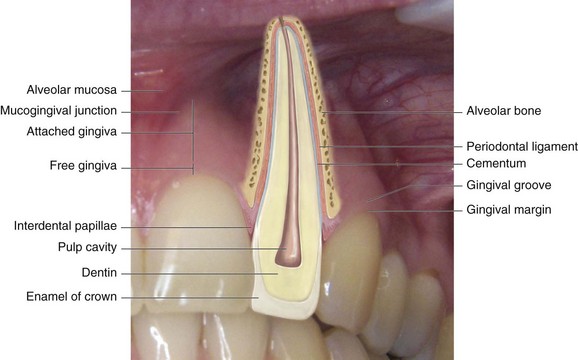
Dentoalveolar traumatic injuries affect many tissues and structures, and the lesion of the affected structures determines the resulting damages. Any prognosis—regardless of whether it is functional or esthetic or involves the pulp and the permanence of the tooth in the oral cavity—requires knowledge of the normal configuration of the tooth and its supporting tissues (Figure 15-1).

Damage can be classified as immediate or delayed. Immediate damages are those that can be observed right after the trauma and involve dislocation of the tissues, more or less complicated dental fractures, laceration of the periodontal ligament, and bone fractures.

Delayed damages are those that manifest themselves later as the sequelae of trauma and should be detected during follow-ups. They include altered root development, periapical infection, and root resorption.

Diagnosis

It is essential to perform a complete first visit of the trauma patient. The patient should be handled calmly but firmly, and the psychological approach should not be underestimated. It is important to realize that one is facing a person who is scared, anxious, and worried about the lesions caused by the trauma and their consequences. As usual, medical history must be collected, along with any existing records and radiographic images, in order to evaluate the presence of predisposing causes for which intervention to prevent further trauma is still possible.

A careful examination must be performed, starting with the extraoral tissues that might have lacerations, followed by examination of the temporomandibular articulation and of the oral cavity, in order to search for any foreign bodies or tooth fragments, evaluate the dental tissues and the position of the teeth, and detect any increase in tooth mobility. The examination continues with palpation of the alveolar process, percussion test of the teeth affected by trauma, and, in the case of dental avulsion, careful examination of the alveoli.

This thorough examination is followed by radiographic imaging (panoramic, intraoral radiographs), photographs, and pulp vitality tests (thermal and electrical).

Treatment

Only after collection of the data will it be possible to plan specific treatment for the type of lesion involved.

The emergency treatment is of utmost importance and should find the dental office prepared, as in some cases it cannot be postponed. Consequently, this may mean canceling appointments with other patients, thus upsetting the office routine. After careful diagnosis, the case is classified and the appropriate emergency treatment can begin. For example, a dental avulsion might require reimplantation and stabilization; pulp exposure might require pulp capping, pulpotomy, or pulpectomy treatment, according to the type of lesion; in the case of crown fracture it is important to find any dental fragments, and luxations require tooth splinting.

Knowledge of pulpal disease is essential to prevent pulp and periapical reactions in the short and long term after the traumatic event. Reversible pulpitis results in the restitutio ad integrum of the pulp, whereas irreversible pulpitis requires pulpectomy and endodontic treatment; necrosis always calls for endodontic treatment.

Crown Fractures

In the case of crown fracture the extent of the lesion must be evaluated. It is important to establish whether the fracture is limited to the enamel and dentin tissues, if the fractured fragment is available, if the pulp is exposed, and if there is pulp necrosis. Palpation makes it possible to assess the mobility of the tooth and alveolar process, and this is followed by an intraoral radiograph to evaluate root formation, and vitality test(s).

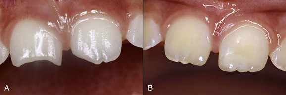
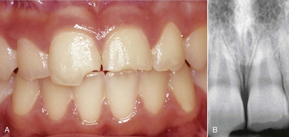
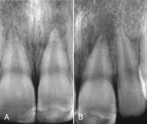
In the case of class A, B, and C coronal fractures (Figures 15-2, 15-3, and 15-4) according to the Spinas-Piroddi classification, treatment involves composite restoration or reattachment of the original fragment when found. Class B1 and C1 coronal fractures (Figures 15-5 to 15-10) likewise require composite restoration or reattachment of the original fragment when found. If root formation is not complete, the treatment entails pulp capping, whereas if the pulp is necrotic the tooth must be treated endodontically. When the pulp is necrotic but the apex is immature, apexification of the root must be induced. Class D and D1 fractures (Figures 15-11 to 15-14) require composite restoration or reattachment of the recovered tooth fragment. If root involvement extends below the alveolar ridge, the treatment requires a clinical crown-lengthening procedure or orthodontic extrusion. If the pulp is affected, the dentist must evaluate whether the tooth should undergo endodontic treatment or a pulp capping procedure.

Stay updated, free dental videos. Join our Telegram channel

Jan 1, 2015 | Posted by in Dental Materials | Comments Off on 15: Traumatology

VIDEdental - Online dental courses

Get VIDEdental app for watching clinical videos