Chapter 10
Mixed Density and Radiopaque Lesions
Aim
It is probably true to say that dentists are more familiar with radiolucencies than they are with radiopacities. The aim of this chapter is to give a brief overview of the common mixed density and radiopaque lesions that can present on intra-oral radiographs. A few examples of the less commonly occurring conditions are included for a more complete overview.
Introduction
As described in Chapter 1, radiopacity is observed when an area, relative to its immediate surroundings, has greater thickness, a higher mean atomic number or a greater physical density. Teeth are the commonest radiopacities on dental radiographs, and anomalies of dental development (e.g. supernumerary teeth) should always be considered first in diagnosis. There are, however, numerous other causes and we have chosen to concentrate upon the most common or most significant lesions here. To study mixed density and radiopaque jaw lesions in detail, consult the further reading section at the end of this chapter. We have already presented some conditions and lesions that fall into this category and when appropriate we cross-refer to the appropriate chapter rather than repeat information.
Choice of Radiographs
This is identical to the situation with radiolucent lesions. Periapical radiography remains the first choice examination of a small, localised lesion and provides optimal image detail. The need for larger radiographs is dictated by size or the presence or likelihood of multiple lesions.
Assessing Mixed Density/Radiopaque Lesions
As with radiolucencies, assessment must take into account a variety of clinical features relating to the patient (age, sex, race, concurrent medical conditions) as well as the radiological signs. Nevertheless, the important radiological signs to consider are listed below.
The site of the lesion
The probability that a lesion is odontogenic in origin is increased if it is found within alveolar bone and, in the mandible, if it lies above the mandibular canal.
The radiopacity of the lesion
This is very important. As a rule of thumb it is useful to look at the radiopacity relative to normal structures such as metal restorations, enamel, cortical bone and trabecular bone (in decreasing order of relative radiopacity). Foreign metallic material, such as retained amalgam, is easy to spot because of its extremely high radiopacity. If a lesion resembles enamel in density, it is suggestive that it does contain enamel i.e. it is an odontome or dental malformation.
The margin of the lesion
In general, although the nature of radiopaque lesions is very different from the radiolucencies described in Chapter 9, there are some similarities. A clear-cut margin suggests a slow, benign growth. A diffuse margin is more suggestive of infection or, extremely rarely, malignancy. A thin radiolucent capsule is a very useful sign as this often indicates a lesion that is odontogenic in origin.
Multiplicity of lesions
Multiple radiopacities are seen in cemento-osseous lesions, multiple osteomas (Gardner’s syndrome) and in “mature” Paget’s disease.
The effect of the lesion on other structures
As with radiolucencies, slow-growing lesions may displace structures such as teeth and the mandibular canal. Resorption may also be seen.
Position
Unlike radiolucencies, radiopacities may lie within the soft tissues and be superimposed upon bone. The differential diagnosis of soft tissue radiopacities is different from that of bony lesions and thus localisation is important.
Mandibular and maxillary tori
The mandibular and maxillary tori are developmental bony overgrowths or exostoses.
Clinical features
Mandibular tori are found in less than 10% of the population and may occur singly, but most often are found bilaterally. They are found on the lingual aspect of the mandible in the premolar region. Tori tend not to develop until middle age when they appear as a painless, lobular bony swelling covered by normal looking mucosa, as shown in Fig 10-1.
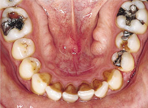
Fig 10-1 Clinical photograph of large bilateral mandibular tori.
The maxillary torus is more common than the mandibular torus, occurring in about 20% of the population. It is found in adults as a midline smooth round or fusiform swelling in the midline of the posterior part of the hard palate. Most are inconspicuous but occasionally can reach a significant size.
Radiological signs
A mandibular torus tends only to show on a radiograph when relatively large. When it does so, it appears as ovoid or circular radiopacity lying close to the alveolar crest (Fig 10-2).
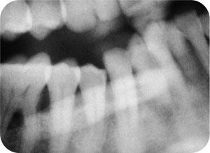
Fig 10-2 Part of panoramic radiograph showing a large mandibular torus superimposed upon the image of the teeth.
A maxillary torus will only show on a radiograph when large. It is only likely to be seen on upper occlusal radiographs or DPRs, when it appears as a well-defined dense radiopacity in the midline of the palate.
Compound odontome
Odontomes are hamartomatous odontogenic entities that consist of varying amounts of enamel, dentine, cementum and pulpal tissue.
Clinical features
These are identified by radiography. This may be a chance discovery or related to the clinical finding of a disturbance in normal tooth eruption. They are most common in the anterior maxilla (62%). While they are commonly identified in children, they will, of course, persist and may not be found until later in life.
Radiological signs
The compound odontome is a 1 to 3 cm-diameter rounded mass of lobulated radiopacities, representing tiny “denticles”, surrounded by a thin radiolucent capsule (Fig 10-3). The enamel in the lesion will make the radiopacity high. As these are “dental” structures, they can be associated with dentigerous cyst formation. While they are associated with malpositioning of adjacent teeth, resorption is not a typical feature.
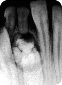
Fig 10-3 A very typical appearance of a compound odontome. The lesion was identified by radiography, carried out because of the retention of 81 and 82. The unerupted 41 partly overlies the odontome. Note the multiple small denticles and the surrounding radiolucent capsule.
Complex odontome
This is rarer than the compound variety of odontome and is characterised by a lack of internal structure. The dental tissues within the odontome are arranged in a more haphazard way.
Clinical features
The likeliest presenting feature is failure of eruption of a tooth or teeth. The most common site is the molar region of the mandible.
Radiological signs
The complex odontome is generally larger than the compound type. It is a densely radiopaque mass without any regular internal structure (Fig 10-4). A radiolucent capsule should be discernible.
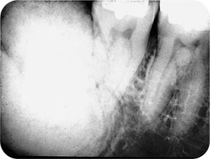
Fig 10-4 A large complex odontome in 48 region. Like the compound variety, the complex odontome has a thin radiolucent capsule. The high radiopacity reflects the enamel content.
Sclerosing osteitis
This inflammatory reaction within bone, typically associated with dental disease, is described in Chapter 5.
Socket sequestrum
Extraction of a tooth may leave fragments of socket wall that are devitalised and that act as sequestra.
Clinical features
It is likely that small fragments of bone are very common and cause no problems. You are most likely to identify one when a patient returns in pain after an extraction. There is likely to be obvious inflammation, debris and pus in the socket and the sequestrum may be visible, or palpable using a probe.
Radiological signs
The sequestrum will be visible as an irregular radiopaque mass overlying the socket, with a line of demarcation separating it from normal bone (Fig 10-5).
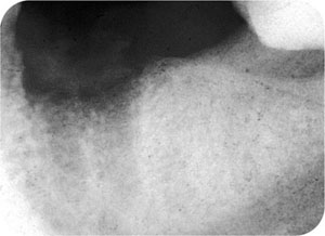
Fig 10-5 Socket sequestrum. This patient had undergone extraction of 36 two weeks previously, but had suffered “infection” in the socket. The sequestrum appears as the irregular radiopacity at the top of the socket, with a thin demarcating line between it and the underlying bone.
Osteomyelitis
See Chapter 5.
Osteosclerosis
Also known as “idiopathic osteosclerosis” or “dense bone island”. This condition commonly causes diagnostic problems for dentists.
Clinical features
There are no clinical signs or symptoms. The abnormality is a chance radiographic finding.
Radiological signs
Usually in the mandibular premolar or molar regions, it appears as a focal area of increased bone density. The shape is irregular and variable, but the margins are sharp. Typically it is found around the root or roots of lower posterior teeth (Fig 10-6). In some cases, the condition is not found against a tooth root, but may be positione/>
Stay updated, free dental videos. Join our Telegram channel
VIDEdental - Online dental courses


