1 Median diastema
Summary
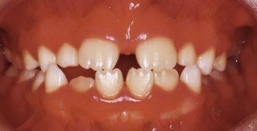
Brian is almost 8 years of age. He presents with a gap between his upper front teeth and crooked lower front teeth (Fig. 1.1). What are the causes of these problems and what treatment would you recommend?
History
Examination
Intraoral examination
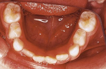
Soft tissues are healthy and the dentition is caries-free. The intraoral views are shown in Figures 1.1 and 1.2.
 erupted but not shown).

 present but
  erupting into
  ).
). ; slight spacing distal of
  .
. flared distally.
 .
.
 rotations?
 What are the possible causes of the upper median diastema?
| Causes | Comments |
|---|---|
| Developmental | Due to pressure of  on on  roots (formerly referred to as ‘ugly duckling’ stage); tends to resolve by the time roots (formerly referred to as ‘ugly duckling’ stage); tends to resolve by the time  erupt erupt |
| Dentoalveolar disproportion | Small teeth in a large arch |
| Absent or peg-shaped 2’s | |
| Supernumerary tooth/teeth in midline | |
Proclination of  |
May be due to digit sucking habit |
| Prominent labial frenum | Implicated where there is blanching of the incisive papilla on stretching the frenum and notching between  is seen in radiograph is seen in radiograph |
| Pathological | Cyst/tumour |
| Juvenile periodontitis |

 may indicate inherent crowding. Also, there is no lower primate (anthropoid) space between the primary canines and first primary molars.
Stay updated, free dental videos. Join our Telegram channel
VIDEdental - Online dental courses


