(b) 
What further details are required?
A detailed history revealed an episode of acute spontaneous pain in the lower right premolar region some nine months previously. This pain had lasted for 3 days; at that stage the patient had not sought dental treatment. He had contacted his general dental practitioner after the cutaneous sinus tract had appeared. The patient complained that the sinus tract was mildly uncomfortable, and he was unable to shave properly. He had no symptoms from any of his teeth.
Medical history
Unremarkable.
Dental history
The patient was an irregular attender, and generally only presented when in discomfort.
Clinical examination
An extraoral sinus tract was visible on the cheek (Figure 1.4.1a).
Intraoral examination revealed that lower right second premolar (45) was mildly tender to percussion. There were normal periodontal probing depths associated with the tooth, however, there was tenderness to palpation adjacent to the apex of 45. The tooth had been restored with a well adapted amalgam restoration.
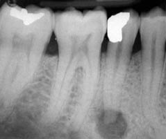
A radiograph revealed a periapical radiolucency associated with the 45 (Figure 1.4.1b). An attempt was made to track the sinus with gutta-percha. This was confounded by the fact that the sinus tract appeared to be narrow and tortuous. The adjacent teeth responded positively to electric pulp testing and cold thermal sensitivity testing.
Diagnosis and treatment planning
What was the diagnosis?
Chronic periapical periodontitis with an extraoral sinus associated with an infected necrotic root canal.
The treatment options discussed with the patient were:
- Root canal treatment.
- Extraction.
- Leave alone.
The root canal appeared to have a simple shape. Therefore, disinfection and obturation would be relatively straightforward; in these cases root canal treatment should have a predictable successful outcome.
Stay updated, free dental videos. Join our Telegram channel

VIDEdental - Online dental courses


