and Angela J. Yoon1
(1)
Columbia University College of Dental Medicine and Department of Pathology and Cell Biology, Columbia University Medical Center, New York, NY, USA
Mucosal lumps and bumps are more accurately referred to as submucosal nodules. In general, submucosal nodules do not spontaneously resolve and often require surgical excision for treatment. A retrospective analysis of 3,129 oral pathology biopsy specimens from pediatric patients (0–18 years of age) submitted for diagnosis to Columbia University Medical Center, Department of Oral and Maxillofacial Pathology found mucocele and fibroma to comprise the two most frequently diagnosed soft tissue lesions. In this chapter, we review the clinical appearance, etiology, differential diagnosis, and recommended treatment for submucosal nodules.
Gingival nodules are covered in Chap. 5.
|
Mucosal lumps and bumps with smooth surfaces
|
|---|
|
Mucocele
|
|
Fibroma
|
|
Pyogenic granuloma
|
|
Hemangioma, vascular malformations, lymphangioma
|
1.1 Mucocele
Clinical Appearance
Smooth-surfaced soft tissue mass or nodule that often has a slight bluish hue. Mucoceles vary in size from a few millimeters to a few centimeters. Some patients might report that the lesion increases and decreases in size. They are often asymptomatic. The surface can appear ulcerated if the child chews or habitually bites it.
Etiology
Trauma resulting in the spillage of mucin into the connective tissues.
Location
Anywhere salivary glands are located; the lower lip is the most common location.
Differential Diagnosis
Salivary duct cyst, salivary gland tumor, fibroma, pyogenic granuloma, benign connective tissue tumor (i.e., lipoma, neuroma, etc.).
Treatment
Surgical excision of the lesion together with adjacent minor salivary gland lobules. It is important to inform the patient and parent/guardian that the lesion can recur if retraumatized (or if offending/damaged salivary gland is not removed). When removing a lower lip mucocele caution should be taken not to nick the labial artery.
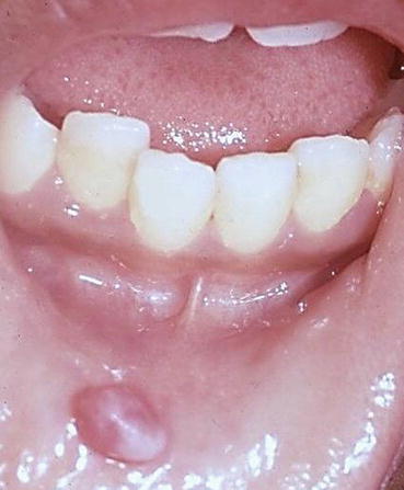
Fig. 1.1
Mucocele. Raised pink nodule of the lower lip
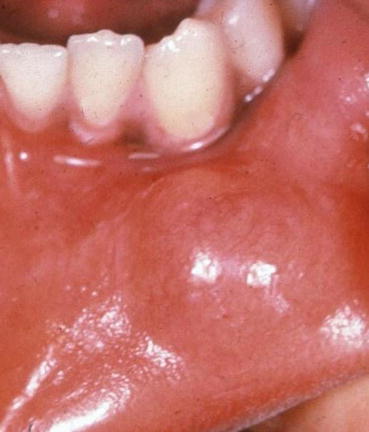
Fig. 1.2
Mucocele. Submucosal nodule of the lower lip
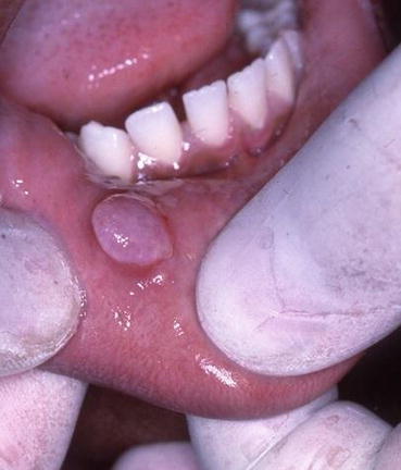
Fig. 1.3
Mucocele. Raised pink, semi-translucent nodule of the lower lip
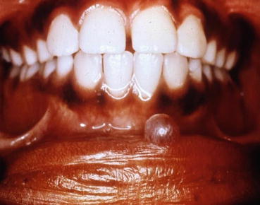
Fig. 1.4
Mucocele. Raised semi-translucent nodule of the lower lip
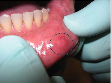
Fig. 1.5
Mucocele. Submucosal nodule of lower lip, best appreciated by palpation
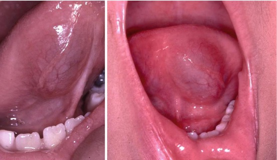
Figs. 1.6 and 1.7
Mucoceles. Submucosal nodules of the ventral tongue. A faint bluish hue can be appreciated
Note
Surgical removal of a mucocele of the tongue often requires deeper dissection since the damaged salivary glands are often situated within the skeletal muscle layer.
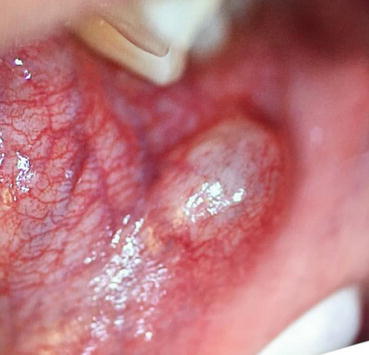
Fig. 1.8
Mucocele. Raised submucosal nodule of the buccal mucosa at the occlusal plane
1.2 Ranula
A ranula is a mucocele of the floor of the mouth involving the sublingual gland or possibly the submandibular gland (less often). Ranulas are most common in children and young adults.
Clinical Appearance
Smooth-surfaced soft tissue mass/swelling occurring on the floor of the mouth, lateral to midline. Ranulas often have a slight bluish hue and can reach several centimeters. The lesion can increase and decrease in size. The child might complain of discomfort or can be asymptomatic.
Etiology
Trauma or obstruction of one of the ducts of the sublingual gland or of the submandibular duct.
Location
Floor of mouth, lateral to midline.
Differential Diagnosis
Salivary gland tumor, dermoid cyst, and abscess.
Treatment
If caused by obstruction from a sialolith (salivary duct stone), removal of the stone can be curative. Otherwise, surgical excision of ranula together with the feeding sublingual gland is the definitive treatment. A less invasive approach is to de-roof the ranula and allow the tissue to granulate. However, with this approach, the lesion is more likely to recur.
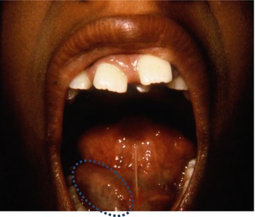
Fig. 1.9
Ranula. Fluctuant submucosal mass of the right floor of mouth with a faint bluish hue
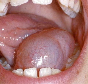
Fig. 1.10
Ranula. Fluctuant submucosal mass of the left floor of mouth causing tongue displacement
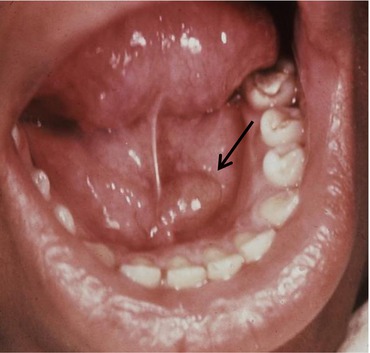
Fig. 1.11
Ranula. Submucosal mass of the left floor of mouth
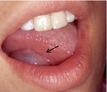
Fig. 1.12
Ranula. Bluish submucosal mass of the right floor of mouth


