Abstract
To ensure that orthognathic surgery is successful, functional aspects such as mastication, pronunciation, swallowing and aesthetic factors must be considered. For successful orthognathic surgery, the orthodontist and the surgeon must constantly study and discuss accurate facial analysis, presurgical orthodontics, choice of appropriate surgical methods, and postsurgical orthodontics.
In this article, we will discuss the team approach for successful orthodontic treatment and orthognathic surgery, establishing close cooperation between the orthodontist and surgeon.
Introduction
Skeletal anchorage devices such as bone plates and miniscrews have expanded orthodontic treatment options compared with those in the past, but orthognathic surgery is still required for adult patients with severe skeletal deformities.
In recent years, orthognathic surgery has improved the effectiveness and stability of treatment. Thanks to improved anesthesia, orthodontic treatment, and surgical methods, there has been an increased demand for active treatment by patients, and more attention is being paid to orthognathic surgery than ever before.
Historically, orthognathic surgery was performed without any presurgical orthodontic treatment and orthodontic treatment was relegated to just postsurgery, if necessary. However, since the 1990s, compensated dentition has been decompensated by orthodontic treatment before surgery after the maxilla and mandibular arches have been coordinated through adjustment. In this way, occlusion can be achieved at the time of the orthognathic surgery, providing improved results and less chance of recurrence.
Recently, the development and advances in cone-beam computed tomography (CBCT), 3D cameras, laser scanning and various computer-aided surgical simulations (CASS) have made the creation of diagnostic and treatment plans for orthognathic surgery much easier and more accurate than in the past ( Fig. 1 ). Furthermore, the development of orthodontic materials has enabled more efficient orthodontic treatment which is of benefit to patients and surgeons alike. In addition, advances in instruments, fixation methods, equipment, and surgical methods for orthognathic surgery have shortened the overall treatment time and improved the postoperative stability.

Now, model surgeries and splint manufacturing have been streamlined by the use of virtual systems ( Fig. 2 ). At the same time, a growing number of patients are electing the “surgery-first” approach to prevent their facial profile from worsening during the presurgical orthodontic phase. While this approach improves their facial profile early in the process, it requires an accurate, well-established prediction of the outcome to minimize any postoperative problems.

For orthognathic surgery to be successful, close coordination between the orthodontist and surgeon is essential and should be based on the initial diagnosis. It may also be necessary to collaborate with prosthodontists, periodontists, plastic surgeons, and other dental specialists, if necessary. This article describes a series of procedures from diagnosis to finishing of orthognathic surgery and examines the collaboration process between orthodontists and surgeons.
Initial evaluation
Patient consultation
Patients who need treatment for malocclusion tend to visit an orthodontist first and want to finish with orthodontic treatment rather than surgery. However, if the patient just wants to improve their appearance, they tend to visit a plastic surgeon or oral and maxillofacial surgeon first. Acknowledging the patient’s specific expectations is important for future treatment planning. The treatment plan will depend on the purpose of the surgery such as to improve a malocclusion, enhance appearance, or both.
Diagnosis
Surgeons set basic aesthetic treatment goals based on patient complaints and on an aesthetic facial examination conducted with the patient’s head in a natural position. Cephalometric measurements and an intraoral exam are used to determine the best type of surgery; single or double jaw surgery and other surgical options (genioplasty, malarplasty, mandibular angle reduction, etc.) for aesthetic purposes.
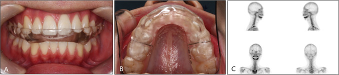
The orthodontist is the one who decides whether orthodontic treatment is necessary when orthognathic surgery is performed. Consideration is given to tooth movement, anchorage preparation, and the need for extractions. A treatment plan is created in consideration of all diagnostic data and patient concerns. Since temporomandibular joint (TMJ) disorder is more frequent in cases with maxillofacial deformity, a TMJ evaluation is necessary with all patients. Pain, joint noise, range of mandibular movement, mandibular displacement upon mouth opening should all be assessed, and referral to a TMJ specialist if additional evaluation or treatment is necessary ( Fig. 3 ).
Surgical treatment objective (STO)
Surgical treatment objective (STO) is the process of performing two-dimensional or three-dimensional virtual orthognathic surgery using lateral cephalograms and CBCT images of patients with a facial skeletal deformity. STO is the basis for diagnosis and planning the treatments of patients who will undergo orthognathic surgery, and STO, based on the prediction of presurgical orthodontic treatment using initial images from lateral cephalograms, is very important as the first step in orthognathic surgical treatment.
Surgeons and orthodontists can predict and evaluate the postoperative stability and aesthetic effects of a treatment with STO. STO also informs the patient of the predicted outcome and serves as the basis of dialogue between the patient and treatment team in the preoperative planning stage ( Fig. 4 ). In other words, the direction and goals of presurgical orthodontic treatment can be set through STO, and it is possible to make a plan for functionally and aesthetically successful orthognathic surgery by predicting the changes in facial profile after the maxillomandibular complex has been surgically moved.

Presurgical orthodontic treatment
The goal of presurgical orthodontic treatment is to arrange maxillary and mandibular teeth in each arch precisely so that the dentition and arches are well coordinated at the time of surgery, to move the maxillary and mandibular incisors to the most appropriate anterior, posterior or vertical position, and to adjust arch width for good occlusion.
Most orthognathic surgery patients show dental compensation. Therefore, the inclination of maxillary and mandibular incisors should be fully decompensated during the presurgical orthodontic phase, and then the angle and anteroposterior relation between the cranial base and maxillary occlusal plane are determined through surgery to carry out sufficient improvement of the soft tissue profile. However, in this case the maxillary incisors were proclined during presurgical orthodontics due to anterior crowding and we decided to improve the torque during Le Fort I surgery ( Figs. 5–8 and Table 1 ).


| Measurement | Norm | Pretreatment | Presurgical treatment | Posttreatment |
|---|---|---|---|---|
| SNA (°) | 82.0 | 85.4 | 85.4 | 84.8 |
| SNB (°) | 80.0 | 87.5 | 86.5 | 82.1 |
| ANB (°) | 2.0 | −2.1 | −1.1 | 2.7 |
| Wits (mm) | 0.0 | −13.2 | −11.2 | −4.0 |
| SN-MP (°) | 32.0 | 38.1 | 39.3 | 39.6 |
| Ramus Height (mm) | 44.0 | 45.1 | 44.7 | 41.4 |
| LFH (ANS-Me/N-Me) (%) | 55.0 | 55.4 | 55.5 | 54.0 |
| U1-SN (°) | 104.0 | 112.8 | 120.0 | 111.2 |
| U1-NA (°) | 22.0 | 27.4 | 34.7 | 26.3 |
| U1-NA (mm) | 4.0 | 6.9 | 7.7 | 4.6 |
| IMPA (°) | 90.0 | 76.5 | 85.5 | 87.1 |
| L1-NB (°) | 25.0 | 23.2 | 32.1 | 32.2 |
| L1-NB (mm) | 4.0 | 6.2 | 8.1 | 6.4 |
| U1/L1 (°) | 131.0 | 131.5 | 114.3 | 118.7 |
| UL-E (mm) | −4.0 | −4.3 | −3.6 | −3.2 |
| LL-E (mm) | −2.0 | 0.3 | 1.5 | −1.2 |
Stay updated, free dental videos. Join our Telegram channel
VIDEdental - Online dental courses


