Key points
- •
Assessing the quality and quantity of the skin is paramount to achieving good results.
- •
Short scar facelifts can be performed in any plane (deep plane, SMAS, subcutaneous).
- •
Adjunct procedures help improve all types of facelifts (ie, skin resurfacing or volume enhancement due to deflation).
- •
Similar complications exist with short scar facelifts and traditional techniques.
- •
Learn the art of facial cosmetic surgery by understanding descent, deterioration and deflation of the aging face.
Short scar facelift techniques have gained popularity over the past 15 years for patients who seek facial rejuvenation. The advantage of reduced morbidity, shorter scars, and less downtime is very appealing for both the patient and the facial cosmetic surgeon. In my opinion, the short scar facelift is the most contemporary facelift and the facelift of the twenty-first century. Understanding the fundamental principles of facial rejuvenation is essential to achieving good results with any facelift technique but is even more essential in achieving good results with short scar facelift techniques.
A common misconception about short scar facelifts is that because the incision is short, then the amount of flap dissection must be short to. When a short scar facelift is performed properly, the amount of dissection is the same as a classic facelift except the posterior incision and the most posterior dissection of the posterior neck is eliminated. Dissection of the posterior neck can still be accomplished from the retroauricular sulcus incision. Dissection in the submental and lateral areas of the face, as well as plication of the anterior and posterior platysma, are still the same as in a classic facelift. Because of the smaller incisions, exposure of the underlying tissue in short scar rhytidectomy is technically more difficult than a classic facelift and it can be frustrating to manipulate the superficial musculoaponeurotic system (SMAS) layer and to achieve hemostasis.
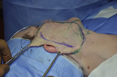
Another misconception about short scar facelifts is that because the incision is shorter, then the amount of skin removed must also be less. Actually the amount of skin removed is essentially the same as a classic facelift except no skin or hair is removed in the occipital area. In the temporal and preauricular areas and the area of skin under the ear lobule, the same amount of tissue is removed for both types of facelifts ( Fig. 1 ).
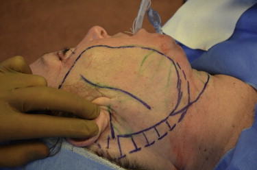
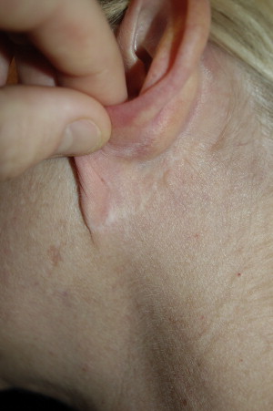
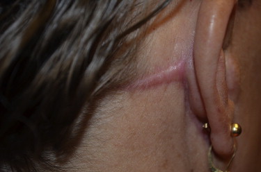
Technically, a short scar remains a short scar if the incision remains in the retroauricular sulcus. Different types of retroauricular incision designs exist. Some techniques include a retroauricular sulcus incision and some techniques do not include a retroauricular incision at all. After performing nearly 1800 short scar facelifts I find it beneficial to drop the incision in the retroauricular sulcus described by Brandy ( Fig. 2 ). The advantage of this small extension is that it helps tighten the lower neck tissue and improves treatment in patients with more severe cervical skin laxity. It also allows better access to the posterior platysma and makes it easier to deal with dog ears in the retroauricular area. Initially, I thought dropping the incision in this area might have negative consequences of poor scarring or visibility. After evaluating more than 1800 of these incisions I can confidently tell you that, when done properly, the incision heals without being noticeable and rarely forms a hypertrophic or keloid scar ( Fig. 3 ). On the contrary, the classic facelift incision that involves the posterior temporal hairline can often heal with a distortion of the hairline with a widening or hypertrophy of the scar ( Fig. 4 ).
Suspension sutures used in short scar rhytidectomy
One of the major differences in some short scar facelift techniques is the use of purse-string sutures. Purse-string sutures for the purpose of lifting the SMAS was introduced by Saylan in 1999. Multiple other purse-string suture techniques have then since been introduced. Purse-string sutures create multiple microimbrications of the SMAS tissue. This leads to a stable volumetric shift of the subcutaneous tissue of the face. Purse-string sutures are typically anchored to a stable point on the face. The overlying periosteum of the zygomatic arch and the deep temporal fascia have been described as anchoring points in the S-Lift, QuickLift, and the Minimal Access Cranial Suspension lift (MACS lift). The suture used for the purse-string can be nonresorbable or resorbable. Purse-string sutures create a powerful upward suspension of the desired aging tissue. Although no great studies have shown the impact of the purse-string suture on the SMAS over time, most likely the scarring formed by the overlying subcutaneous tissue and the fibrous adhesions between the microimbrications holds the newly suspended SMAS tissue in place. This presents one of the problems with suspension sutures. The use of permanent sutures can cause palpability of knots and the potential for long-term infection. Knowing that the face deflates and thins with time, it only makes sense that the probability of these sutures having problems after 10 to 20 years is high. Suspension suture techniques tend to be used on younger patients receiving facelift and time will tell if we see more problems with nonresorbable purse-string sutures. On the other hand, resorbable sutures also can be used. The problem with resorbable sutures is that the ideal materials, either polyglycolide or polydioxanone, should not be used for tissues requiring prolonged tension. Polyglycolide has only 23% of its tensile strength at 4 weeks and polydioxanone has 44% at 8 weeks. The question is how long do resorbable sutures need to maintain tensile strength to prevent movement of the SMAS tissue when used as a suspension suture in facelifts? When suspension sutures in the face are placed, a moderate amount of tension exists in the gathering and lifting of the SMAS layer and the resorbable suture is losing strength rather quickly. The ideal suture would retain 100% tensile strength for approximately 90 days and then completely degrade after the newly formed fibrous tissue holds the suspended SMAS tissue in place. I have removed multiple permanent purse-string sutures months after their placement and have found the SMAS tissue and skin to remain stable after removal.
Another problem encountered with purse-string sutures is the gathering of tissue as the purse-string is tightened. As the suture is tightened, the tissue moves upward but also inward and outward, entrapping any tissue within the circumference of the purse-string. Most commonly this tissue needs to be removed and sculptured to prevent lumps and bumps of the tissue. If the purse-strings go outside the area of the underlying parotid gland, then the suture has the ability to also entrap branches of the facial nerve and cause paralysis. The purse-strings also have the potential to cause lymphatic congestion and can prolong the swelling under the surgical flaps. I have seen both of these complications; fortunately, the complications are rare and if a facial nerve paralysis occurs, the treatment is removal of the offending purse-string with potential for recovery. Tonnard and colleagues also have reported prolonged swelling and facial edema in the area of the purse-strings for up to 17 weeks.
The three “Ds” of facial aging
The quality and quantity of the skin helps determine the correct type of facelift procedure for each individual patient. If the patient has a significant excess of skin with poor elasticity, the decision to create longer incisions must be contemplated ( Fig. 5 ). The ideal patient for a short scar technique should have mild to moderate skin laxity and good to excellent skin quality ( Fig. 6 ).
Another very important aspect of facial evaluation is determining what I call the 3 “Ds” of facial rejuvenation ( Table 1 ).
| Deterioration | Deflation | Descent |
|---|---|---|
| Glogau Classification: I, Mild: Little wrinkling II, Moderate: Early wrinkling III, Advanced: Wrinkling at rest IV, Severe: Wrinkling at rest with associated cutis laxis |
I, Mild: Small prejowl sulcus, slight hollowing of temporal, periorbital, and cheek areas II, Moderate: Deeper prejowl sulcus, moderate hollowing of temporal, periorbital, and cheek areas III, Severe: Periorbital, perioral, temporal and cheek hollowing |
I, Mild: Early jowls, slight cervical skin laxity, minimal to no platysmal bands II, Moderate: Moderate jowls, moderate cervical laxity, minimal platysmal bands III, Severe: Heavy jowls, active platysmal bands, significant cervical laxity or large obese necks |
The first “D” stands for deterioration of the skin. Deterioration of the skin can be explained by the Glogau Classification (see Table 1 ). Patients with mild to moderate Glogau Classifications are the best candidates for short scar facelift techniques. To achieve optimal results, patients with advanced and severe Glogau Classifications should have some type of aggressive facial resurfacing procedure before any facelift technique. Patients having any type of facelift procedure with a severe Glogau Classification, without first addressing the real problem, which is the skin quality, are destined for failure. Optimally, these patients should have either full-face deep CO2 or deep dermabrasion at least 3 months before any facelift procedure ( Fig. 7 ). I have not seen any real dramatic results with the new fractional or radiofrequency lasers. If anything, I get many unhappy patients who have had those therapies and they come to see me for correction of their still sagging jowls and neck.
The second “D” stands for deflation. Deflation of the aging face has been well documented. Dr Val Lambros studies suggest “that gravity’s role in aging has been exaggerated, and that the downward migration of facial skin over time is merely an illusion. The real culprit, the study found, is the loss of volume underneath the skin.” Regardless, if the real culprit is deflation, you should never overlook the first “D” for deterioration of the skin. Giving a patient volume without restoring the quality of the skin first is a recipe for disaster, especially if the patient’s primary reason for facial aging is deterioration. Deflation can be apparent with temporal hollowing and sunken periorbital areas and the cheeks ( Fig. 8 ). Volume replacement can be achieved with alloplastic implants, fat grafting, or some kind of filler.
The third and final “D” stands for descent. Descent of the facial skin can be seen in the brows, nasolabial folds, jowls, and especially in the submental area and lower neck ( Fig. 9 ). The key here is that in most cases, I believe descent of facial tissue is really a problem of the first two “Ds.” If the primary problem of the aging facial patient is poor skin quality or deterioration of the skin, then you will see descent of the tissue. Likewise, if the main problem of the aging facial patient is deflation, you can also see descent of the tissues.
In my opinion, the art of facial cosmetic surgery is learning to decipher what percentage of each of these 3 “Ds” your patient has before you decide to perform any type of facelift procedure. After you have decided on the amount of each of the 3 different “D” percentages of each aging face, you can then come up with a treatment plan. For example, a patient will present to your office for a facelift, when in reality all the patient really may need is an aggressive resurfacing procedure and volume modification ( Fig. 10 ). In this particular situation, I would look at the patient’s face and say “60% of her aging is deterioration of the skin, 30% of her aging is deflation, and maybe only 10% of her problem is descent.” Her treatment plan consisted of a first-stage surgery with deep full-face CO2 and approximately 30 mL of harvested lateral thigh fat grafted to her face. The second stage would be 3 months later, with a lower face and neck lift with possible relasering of stubborn wrinkles and a second fat-grafting session to the face. She was so happy with her first-stage results that she never came back for her facelift. Only after or possibly at the same time, should some patients have a facelift until the other 2 “Ds” have been treated. Most of all my patients with short scar facelift receive some form of resurfacing and volume procedure either before or at the same time as the short scar facelift. Rarely will I perform a short scar facelift by itself. It is for this reason I believe my short scar facelifts have such a high satisfaction rate. A study I performed and presented at the American Academy of Cosmetic Surgery Annual Meeting in January of 2010 showed a short scar facelift 5-year satisfaction rate of higher then 90%. These results were compared with Owsley’s satisfaction rates at 5 years for a traditional type of facelift and were found to be statistically very similar.


