The complexity of a bilateral cleft lip repair must be well understood by any surgeon performing this procedure. Multiple factors play a role in the difficulty that one must overcome to correct the obvious facial deformity. These include a widely displaced lateral lip segment, lack of developed lip tissue in the anterior segment, and a displaced premaxillary segment. All three need to be taken into consideration to obtain an optimal result.
Historically, techniques used for repair of a bilateral cleft lip were based on the repair of a unilateral cleft lip. The procedure was done as a staged process, first repairing the more severe side and correcting the contralateral side at a later date. Following this idea, most procedures were based on the proposal that the prolabium had limited growth potential. This led to multiple designs and geometric shapes to correct for this growth potential. The triangle and rectangle flaps were examples of this idea, creating an abnormally long lip with irregular scars. Soon after, the straight line closure and variants were developed to decrease the unnatural length of the lip; unfortunately, these techniques created problems with aberrant scaring. Not much thought went into the relevance of the orbicularis oris until the latter part of the twentieth century. Others reported on the reconstruction of the muscle, creating continuity between the two lateral lip elements. That procedure was referred to as the functional cleft lip repair. It did have its opponents, however, who thought it would lead to growth restriction of the premaxilla.
Additions stemming from the work of Millard, Mulliken, McComb, Trott, Mohan, and Cutting discuss the correction of the cleft nasal deformity in addition to the repair of the lip. Although extensively described and used by surgeons, the authors believe that the results of the cleft nasal deformity are easier to predict and more stable if the correction is undertaken once most of the child’s growth has taken place.
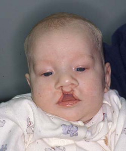
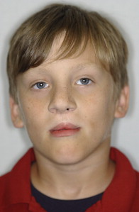
Although not all clefts are similar, the application of basic principles with slight dimensional modifications to the technique generally produces an aesthetically pleasing result ( Figs. 1 and 2 ). The authors’ preferred timing for repair is between the ages of 3 and 6 months.
Surgical technique
The patient is placed in a supine position on the operating table with a small shoulder roll for support. A broad-spectrum antibiotic, typically a cephalosporin, is given before surgery as prophylaxis. Before the procedure, for reduction of the postoperative inflammatory phase, a combination of short- and long-acting steroids is administered unless contraindicated. After induction, an oral endotracheal tube is secured to the patient’s chin in the midline. After surgical preparation, the anatomic structures are palpated and marked with a sterile surgical pen.
Design
Anterior Lip
The design of the planned surgical incisions is marked, starting with the prolabium ( Fig. 3 A ). At the level of the lip-columellar crease in the midline, two points are placed approximately 2 to 2.5 mm apart. The length of the philtral column is then established by marking a point 6 mm inferior to the lip-columellar crease. The widest point on the philtral column, established 1.5 mm superior to the inferior point, is 3 to 3.5 mm in width and forms the peaks of Cupid’s bow. These points are connected to produce the final design of the philtral column (see Fig. 3 B, C). The width of Cupid’s bow and the length of the philtral column are directly related to the age of the patient. Repairs in older patients have less of a tendency to widen over time. The measurements used to design the philtral column should be slightly enlarged in older patients to compensate for this decreased amount of growth potential.
Lateral Lip
The nasal base incisions are first marked bilaterally, creating a curvilinear line along the alar crease. This forms the releasing incisions that aid in the advancement of the lateral lip elements. The peaks of Cupid’s bow are then marked and positioned at the vermilion-cutaneous border, where the vermilion border of the lip and the white roll begin to converge. The marks may then be adjusted to create an equal distance bilaterally from the commissure of the mouth to the marked points ( Fig. 4 ). A line perpendicular to the tangential line at the vermilion-cutaneous border is then marked through the vermilion. After markings, bilateral infraorbital nerve blocks are performed and the anterior and lateral lips and alar bases are injected with 1% lidocaine and a 1:100,000 epinephrine mixture.
Incision
After placement of the moistened throat pack, the first incision is initiated on the prolabium. This incision includes skin and subcutaneous tissue. The flap should be raised from the philtral notch, elevating superiorly and gradually enlarging the thickness of the flap so as to preserve the columellar blood supply. The remaining vermillion and skin are reflected and turned intraorally as a flap based on the premaxilla ( Fig. 5 ). This tissue aids in the intraoral closure of the mucosal surface.
The alar bases are then freed from the lateral lip elements along the curvilinear line ( Fig. 6 ). The vermilion border is incised in a partial-thickness fashion to the point marking the peak of Cupid’s bow. Preservation of the lateral vermilion mucosal flaps inferior to the peak of Cupid’s bow is essential for reconstruction of the central lip region. The dissection to release the lateral lip elements from the maxilla is done with sharp scissors in a submucosal plane. The lateral lip elements must be completely separated from the intraoral mucosa. This dissection is usually performed at the malar prominences for adequate mobilization. The orbicularis oris is separated from the lateral lip flaps in the subdermal plane ( Fig. 7 ). The muscle bundles are separated from the anterior maxilla at the alar base to reorient the fibers in a horizontal direction ( Fig. 8 ). The nasovestibular web is released from its attachment at the piriform rim ( Fig. 9 ). This release allows advancement in an anteromedial direction, correcting alar base width.


