Introduction
In this research project, we aimed to compare the effectiveness of single (1 deciduous canine) and double (deciduous canine and first molar) extractions in subjects with retained maxillary permanent canines positioned palatally or centrally in the alveolar crest, at risk for root resorption of adjacent permanent teeth.
Methods
Subjects at risk for canine impaction or resorptive situations were randomly assigned to 1 of 2 treatment modalities: single extraction (17 patients, 28 canines) or double extraction (20 patients, 37 canines). Thirty-one patients with 53 canines judged to be not at risk constituted the untreated control group. Panoramic radiographs were taken at the initial observation and after 18 months on average. Between-group statistical comparisons were carried out on the changes in canine inclination and sector location (measured on panoramic radiographs) and on the percentages of successful permanent canine eruptions.
Results
The double-extraction group showed significant improvements in the success rate and the intrabony position of the permanent canine, in terms of uprighting the canine’s long axis with a crown movement in a distal direction.
Conclusions
Concomitant deciduous canine and first molar extractions proved to be more effective as a preventive approach to promote eruption of retained maxillary permanent canines positioned palatally or centrally.
Maxillary canine impaction is often encountered in orthodontic clinical practice ; the frequency ranges from 1.7% in the general population to 4.3% in the population of subjects referred to oral surgery or orthodontics departments.
Ectopically or nonerupting canines can lead to resorption of the roots of the adjacent permanent teeth. For this reason, great emphasis should be given to the early detection of ectopic eruption and potential resorptive situations, when preventive measures could reduce the severity of the impaction and, if possible, encourage the eruption of the canine, thus avoiding possible detrimental effects.
For early diagnosis of ectopically erupting maxillary canines, both clinical (digital palpation screening method) and radiographic (eruption angle and position measured on panoramic radiograph) examinations should be used.
Extraction of the corresponding deciduous canines has been recommended as a preventive treatment to promote the eruption of malposed canines. Previous studies found that between 50% and 78% of palatally displaced maxillary canines reverted to a normal eruption path after this procedure. The additional use of headgear to maintain space in the maxillary dental arch resulted in an increase in terms of successful eruption of up to 80% of the canines, or 87.5% of the subjects treated, with a significant improvement in the intraosseous canine position.
The purpose of this study was to evaluate the effectiveness of concomitant extraction of the deciduous canine and first molar as a preventive procedure for corresponding retained maxillary permanent canines positioned palatally or centrally in the alveolar crest, compared with extraction of only the deciduous canine. The outcome of this preventive measure was evaluated in terms of improvement of the intraosseous position of the displaced canine and successful eruption.
Material and methods
In this randomized clinical trial, we analyzed patient records collected from the Department of Orthodontics, University of Bologna, in Italy.
Inclusion criteria were nonorthodontic patients, white ancestry, age between 8 and 13 years, maxillary deciduous canines and first molars in the dental arch, and good-quality panoramic radiographs. Exclusion criteria were previous orthodontic treatment, premature loss of the maxillary deciduous canines and first molars, labially retained maxillary permanent canines, aplasia or severe hypoplasia of the crown of the maxillary permanent lateral incisors, craniofacial syndromes, odontomas, cysts, cleft lip or palate (or both), evidence of traumatic injuries to the permanent incisors or to the face, and multiple or advanced caries. Seventy-one subjects, with 123 canines, fulfilled all criteria and were included in this study, after informed consent was obtained from them or their parents or guardians. All the patients were examined identically, both clinically and radiographically.
Because we considered it unethical not to treat patients at risk for ectopically erupting canines or potentially resorptive situations, canines were diagnosed to be at risk by at least 1 of the following clinical and radiographic criteria, which are widely accepted in the international literature and among practitioners. The clinical criteria were absence of palpation of the canine bulge, canine bulge palpable palatally, and no abnormal inclination or rotation of the adjacent lateral incisor crown. The radiographic criteria were inclination of the canine to a vertical line passing through the midline exceeding 25° and overlapping of the canine crown with the root of the permanent lateral incisor.
Among the 71 subjects (123 canines), 31 patients (53 canines) were judged to be at no risk. They had no treatment and constituted the control group (CG).
The remaining 40 patients (70 canines), diagnosed as at risk, were randomly assigned to 1 of 2 groups: ECG (20 patients, 33 canines), having extraction of only the deciduous canine corresponding to the ectopically erupting maxillary permanent canine; and ECMG (20 patients, 37 canines), having concomitant extraction of the deciduous canine and first molar corresponding to the ectopically erupting maxillary permanent canine.
Randomization was carried out by using a block design and computer-generated random numbers. The allocations were concealed in consecutively numbered, sealed envelopes.
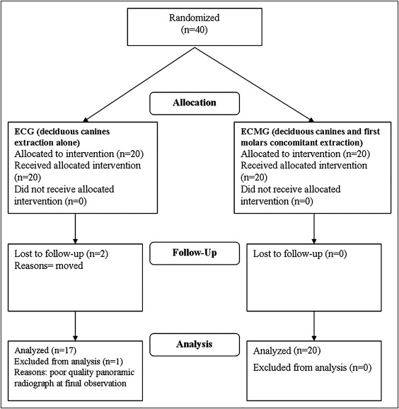
Three patients (5 canines) in the ECG did not complete the clinical trial: 2 were lost to follow-up because they moved, and 1 was not analyzed because of a poor-quality panoramic radiograph at follow-up. Consequently, the final ECG consisted of 17 patients and 28 canines. A CONSORT flow chart of participants in the ECG and the ECMG through each stage of the trial is shown in Figure 1 . Patient distribution by sex and age is shown in Table I .
| ECG | ECMG | CG | Total | |
|---|---|---|---|---|
| Subjects (n) | 17 | 20 | 31 | 68 |
| Male | 9 | 9 | 16 | 34 |
| Female | 8 | 11 | 15 | 34 |
| Mean age (y) | 9.8 | 10.2 | 9.0 |
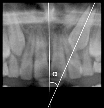
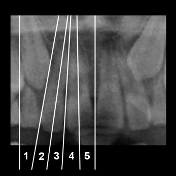
Panoramic radiographs were taken at initial observation (T0) and after an average period of 18 months (T1)—a duration that has already proven to be appropriate. No subject in either treated group received any additional orthodontic or surgical therapy beyond the extraction of the deciduous canine (ECG) or the concomitant extraction of the deciduous canine and first molar (ECMG) between T0 and T1. For all patients, the panoramic radiographs were taken with the same radiologic apparatus at both T0 and T1, under standardized conditions. Since different studies have already confirmed the reliability of angular measurements in panoramic radiographs, each panoramic radiograph was digitized with a scanner (Expression 1680 Pro, Epson, Cinisello Balsamo, Milano, Italy), and the angular values were calculated with measurement software (LightningPlant 1.0.0, ElleSoft, Chieti, Italy). Two radiographic parameters were analyzed to assess the canine eruption pattern: (1) the mesial inclination of the crown to the midline, according to Ericson and Kurol (angle α, Fig 2 ); and (2) the medial crown position in sectors 1-5, according to Ericson and Kurol (s1-s5, Fig 3 ). Radiographic measurements were made at both T0 and T1 by the same operator (S.I.P.), who underwent an intraexaminer reliability check. Additionally, the development of the permanent canine (measuring the length of the root) was evaluated at T0 according to 2 stages, according to the method of Ericson and Kurol : (1) the root was longer that the canine crown, and (2) the root was shorter that the canine crown ( Table II ).
| ECG | ECMG | CG | |
|---|---|---|---|
| Canines (n) | 28 | 37 | 53 |
| Stage 1 | 17 | 20 | 15 |
| Stage 2 | 11 | 17 | 38 |
The between-group statistical comparison was carried out on the T0-T1 changes in the radiographic measurements. The successful outcome was defined, according to Leonardi et al, as the complete eruption of the permanent canine into the dental arch within 48 months from the initial observation, thus permitting bracket positioning for final arch alignment when needed.
Statistical analysis
On the basis of the results obtained in a pilot sampling of canines, a normal distribution of the main variable—the absolute difference of the α-angle of the canine—was hypothesized; similarly, a standard deviation of 13° was estimated. A minimum of 26 canines was required for each group, when the true value of the absolute difference of the α-angle between the ECG and the ECMG was 10°, at an α-level of 0.05 and with a power of 80%.
The Kolmogorov-Smirnov test with the Lilliefors level of significance was carried out to verify the normality of the distribution of the α-angle of the canines in each group. In addition, skewness and kurtosis coefficients (± SE) and cumulative normal plots were used to confirm the Gaussian form of distribution of the α-angles. A generalized linear model was applied to verify the significance of the differences of the α-angles. The values of the α-angles among the 3 groups at T0 were always significantly different. Consequently, to control for the influence of the severity of canine displacement on the final results, the inclination at T0 was used as the covariate; also, the stage of root development of the permanent canines was controlled for. The chi-square test was used to highlight an association of a group with variations of sector. The t test for independent samples was used for comparison between each pair of groups. The Fisher exact test was carried out to compare the percentages of favorable outcomes between the ECG and the ECMG.
An α-level of 0.05 was set and adjusted by the Bonferroni correction at 0.016 for multiple comparisons.
To evaluate the method error, an intraobserver reliability check was carried out for both angular values and sectors. Fifteen randomly selected panoramic radiographs (15 subjects; 30 canines) were measured by the same operator (S.I.P.) twice, on 2 days, with 15 days separating the measurement sessions. The intraclass correlation coefficient (ICC) was 0.968 (95% CI, 0.934-0.985; P = 0.0001) for angulation of the canines, thus confirming high intraobserver reliability for the method used. The ICC was 1.00 for the sector designation, indicating no differences between measurement sessions. To evaluate whether clinical experience affects the accuracy of the measurements, 20 randomly selected panoramic radiographs (20 subjects; 20 right and 20 left canines) were independently measured by 5 orthodontists. The ICC values were 0.955 for the angular measurements and 1.000 for the sector designation for both right and left canines. These coefficients show high interobserver reliability of the measurement method, which has proved not to be influenced by clinical experience.
Stay updated, free dental videos. Join our Telegram channel
VIDEdental - Online dental courses


