A patient came with left-side temporomandibular arthralgia, limited mandibular opening, frontal facial asymmetry, and a significant anterolateral open bite. Severe alterations in the occlusal and maxillofacial anatomy resulted from an osteochondroma associated with the mandibular condyle. We describe the changes associated with extirpation of the mandibular condylar osteochondroma and subsequent orthodontic treatment. These clinical changes resulted in improved facial symmetry and a satisfactory functional occlusion.
Highlights
- •
Osteoma/osteochondroma of the condyle may cause a slow, progressive shift in occlusion, with deviation of the midline of the chin toward the unaffected side.
- •
After osteochondroma extirpation, the mandible rotated clockwise by about 6° on average on both sides and deviated by about 2.5 mm on the left side compared with the preoperative position.
- •
An effective orthodontic treatment regimen can be designed and applied postoperatively through an initial preoperative evaluation of mandibular change.
- •
The preoperative orthodontic treatment should be performed effectively to prevent the malocclusion postoperatively.
Tumors of the mandibular condyle such as osteoma or osteochondroma are rare and grow slowly. Patients may often complain of symptoms such as mandibular deviation and limited mouth opening. These symptoms are treated by extirpation (condylectomy), but after the surgery the occlusion often changes. It has been reported that a Class III malocclusion and anterior open bite can occur after removal of a condylar tumor. However, there have been no clinical case reports describing the preoperative and postoperative occlusal and maxillofacial changes associated with surgical and orthodontic intervention.
In this case report, we describe the occlusal and maxillofacial changes associated with surgical and orthodontic treatment.
Diagnosis and etiology
A woman aged 24 years 11 months came with left-side temporomandibular joint (TMJ) arthralgia and limited mouth opening as her chief complaints. She had sharp pain at the left TMJ on mouth opening, and the maximum interincisal opening was consistently recorded at 20 mm, with deviation to the affected side.
The frontal facial photograph showed a remarkable right-side shift of the mandible; the lower lip “rolled up” with mentalis strain ( Fig 1 ). Closure of the mouth was difficult without significant muscle tensing. The 3-dimensional computed tomography images showed that the left ramus was longer than the right, and the mandible was depressed inferiorly on the left.
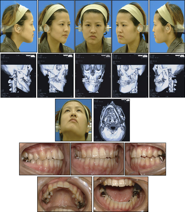
A bilateral Angle Class III malocclusion with an anterolateral open bite was present on the left side ( Fig 1 ).
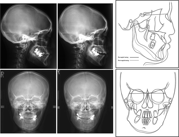
The radiographic findings included a lateral cephalometric analysis ( Fig 2 ; Table ). It showed (1) a Class III skeletal pattern (ANB, −1.7°; SNA, 78.5°; SNB, 80.2°); (2) a mandibular plane angle of 7.1° on the left side, with the right side within the normal range at 29.1°; and (3) maxillary incisors with labial tipping (central incisor to Frankfort horizontal plane, 124.9°); and (4) mandibular incisors with lingual tipping (FMIA, 73.8°).
| Measurement | Preoperation | Pretreatment | Posttreatment | Retention |
|---|---|---|---|---|
| 24 y 11 mo | 25 y 2 mo | 28 y 9 mo | 30 y 11 mo | |
| Lateral | ||||
| SNA (°) | 79.2 | 79.2 | 79.2 | 79.2 |
| SNB (°) | 81.3 | 77.7 | 78.0 | 78.0 |
| ANB (°) | −2.2 | 1.5 | 1.2 | 1.2 |
| FMA (°) (right side) ∗ | 29.0 | 34.2 | 30.8 | 31.4 |
| FMA (°) (left side) ∗ | 7.7 | 22.0 | 28.7 | 31.0 |
| Ramus inclination (°) (right side) ∗ | 18.1 | 9.8 | 11.5 | 7.0 |
| Ramus inclination (°) (left side) ∗ | 4.7 | −6.8 | −5.0 | −4.6 |
| Gonial angle (°) (right side) ∗ | 137.0 | 134.0 | 130.6 | 130.0 |
| Gonial angle (°) (left side) ∗ | 102.4 | 105.1 | 113.7 | 117.0 |
| IMPA (°) (right side) ∗ | 77.5 | 88.2 | 87.0 | 87.5 |
| FMIA (°) (right side) ∗ | 73.5 | 57.6 | 62.2 | 61.1 |
| U1 to FH (°) | 118.0 | 118.0 | 116.0 | 116.0 |
| Occlusal plane angle (°) | 11.3 | 9.6 | 8.8 | 10.2 |
| N-Me (mm) | 121.0 | 126.6 | 124.5 | 125.3 |
| ANS-Me (mm) | 71.0 | 76.2 | 75.2 | 75.5 |
| Posteroanterior | ||||
| Molar relationship, left (mm) | −1.3 | 1.1 | −0.1 | 0.3 |
| Molar relationship, right (mm) | 1.0 | −0.4 | −0.8 | −1.5 |
| Denture midline (mm) | 4.6 | 1.5 | 0.0 | 0.0 |
| Maxillomandibular width, left (mm) | −9.8 | −11.2 | −15.2 | −16.3 |
| Maxillomandibular width, right (mm) | −19.5 | −18.6 | −15.4 | −15.4 |
| Maxillomandibular midline (°) | 10.7 | 8.5 | 1.0 | 0.3 |
| Occlusal plane tilt (mm) | 5.7 | 4.0 | 0.5 | 1.0 |
∗ The right side, the measurement point of the right side (the normal side); the left side, the measurement point of the left side (the affected side).
The frontal cephalometric analysis ( Fig 2 ; Table) showed that the mandible deviated down and to the left side (denture midline, 4.6 mm; maxillary mandibular midline, 10.7°).
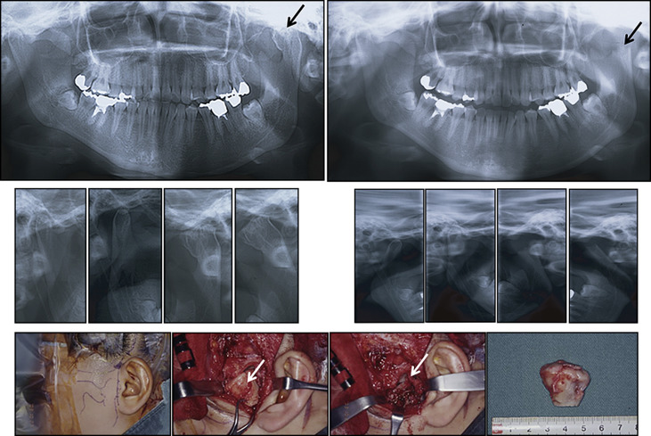
The panoramic radiograph ( Fig 3 ) showed a radiopaque mass associated with the left condylar region of similar density to the adjacent bone.