Introduction
Because of the close anatomic proximity of the maxillary permanent canine and first premolar, we investigated the relationship between their preeruptive positional changes in this retrospective study.
Methods
Sixty-three pairs of panoramic radiographs obtained 12 to 24 months apart from nonorthodontic subjects between 8 and 10 years old were collected. Canine and premolar inclinations to the midline and mesiodistal sector locations were measured, and their changes over time were estimated. The relationship between changes in canine (Δα) and premolar inclinations (Δπ) was explored alone and after adjustment for intraosseous distance between the teeth (S c-p ) and months between radiographs (ΔM).
Results
Except for right canine sector, both canine and premolar inclinations and sectors showed significant differences between time points. Pearson partial correlation analysis showed a strong, positive correlation between Δα and Δπ after adjusting for S c-p and ΔM (right: r = 0.766, P <0.05; left: r = 0.785; P <0.05).
Conclusions
Canine and premolar inclinations and mesiodistal locations varied significantly between 8 and 10 years of age. The strong positive correlation between the preeruptive positional changes of the maxillary canine and the first premolar suggested that with a greater uprighting movement of the long axis of the premolar over time, there was greater uprighting of the long axis of the canine.
Highlights
- •
Canine inclination varied significantly between 8 and 10 years of age.
- •
Premolar inclination and mesiodistal location varied significantly between 8 and 10 years of age.
- •
There is a strong correlation between changes in canine and premolar inclinations.
- •
The more the premolar uprighted over time, the more the canine straightened.
Impaction of the maxillary canine is a challenging problem in orthodontic practice, with a prevalence rate ranging from 1.7% in the general population to 4.3% in patients referred to oral surgery or orthodontic departments. The high prevalence is most likely due to an extended development period as well as to the long and tortuous path of eruption before the canine emerges into full occlusion. Mineralization of the canine begins at 4 to 12 months of age and is complete at 6 to 7 years. At age 8, the canine is high above its predecessor; then it follows a mesial path of eruption until its crown reaches the distal aspect of the lateral incisor root. The erupting canine is gradually uprighted to a more vertical position and is “guided” by the lateral incisor root, until it is fully erupted at mean ages of 10 to 12 years for girls and 11 to 13 years for boys. Palatally displaced canines are reported to occur 3 times more frequently than those found buccally. In case of palatal ectopic maxillary permanent canines, the adoption of suitable preventive measures aimed at redirecting the canines to a normal eruptive path prevents or reduces the occurrence of root resorption of neighboring teeth and canine impaction, thus avoiding a complex and prolonged treatment with an uncertain outcome.
Several preventive measures have been proposed since the 1980s, including early extraction of deciduous canines alone or in association with cervical pull headgear, transpalatal arch, rapid maxillary expansion, or deciduous first molar extraction. Concomitant extraction of a deciduous canine and a first molar (double extraction) is more satisfactory in terms of clinical (ie, spontaneous eruption of ectopic canines) and radiographic (ie, canine inclination to the midline and overlapping with adjacent tooth roots) outcomes compared with the extraction of a deciduous canine alone (single extraction). The finding that double extraction, unlike single extraction, also determines a statistically significant decrease in the permanent first premolar’s inclination led to the hypothesis that a change in premolar inclination might affect the extent of canine uprighting. An understanding of the correlation between the preeruptive positional changes of these teeth should therefore be of critical interest to orthodontists, since it would add a further element to the detection of canines that will most likely present a worsening intraosseous position during their eruption and therefore would benefit from more frequent checkup visits or preventive approaches. However, to date, no studies have tried to determine either the normal maxillary first premolar eruption pattern or its relationship with the canine eruption pattern.
Therefore, in this study we aimed to investigate, in a sample of children not undergoing or not having undergone orthodontic treatment, both the canine and the first premolar physiologic eruptive patterns in the mixed dentition and the relationship between their preeruptive positional changes.
Material and methods
This was designed as a retrospective study. The study sample was derived from the population of patients at the Department of Orthodontics of the University of Bologna in Italy for orthodontic consultations from January 1, 2007, to January 1, 2013. To be eligible for the study, patients had to fulfill the following inclusion criteria: (1) availability in the medical records of 2 panoramic radiographs taken 12 to 24 months apart, (2) white ancestry, (3) age at the time of the first panoramic radiograph between 8 and 10, and (4) maxillary deciduous canines and first molars in the dental arch at the time of the first panoramic radiograph. Patients fulfilling these criteria were informed of the study aims and asked to sign an informed consent to participate. Exclusion criteria were (1) previous or ongoing orthodontic treatment at the time of both panoramic radiographs; (2) aplasia or severe hypoplasia of the crown of the maxillary permanent lateral incisors; (3) craniofacial syndromes, odontomas, cysts, cleft lip or palate (or both); (4) history of traumatic injuries to the face; (5) multiple or advanced caries; and (6) inadequate records.
All data were obtained from manual medical record reviews by 1 operator (D.R.I.). Demographic data and medical and dental histories were collected. Each panoramic radiograph available in the medical records selected for the study was digitized at a resolution of 300 dpi with 256 gray levels with a scanner (Expression 1680 Pro; Epson Italia, Milan, Italy) equipped with a transparency adapter and saved as a JPEG at a quality setting of 8.
Two measurements were performed for both the maxillary permanent canine and the maxillary first premolar to define their intraosseous positions.
- 1.
The angle formed between the long axis of the tooth and the midline (angle α for the maxillary permanent canine, angle π for the maxillary first premolar; Fig 1 ). Angular values were calculated on the digital images using the ImageJ angle measurement tool.
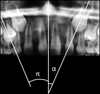
Fig 1 Permanent maxillary canine inclination to the midline ( angle α ) and maxillary first premolar inclination to the midline ( angle π ). - 2.
The mesiodistal position of the tooth. The locations of the canine cusp tip (S c ) and the first premolar bud (S p ) were assessed in accordance with the sector analysis shown in Figure 2 : sector 0, area distal to a line tangent to the distal heights of contour of the deciduous canine crown and root; sector 1: area bounded by sector 0 and the long axis of the deciduous canine; sector 2: area bounded by sector 1 and a line tangent to the distal heights of contour of the lateral incisor crown and root; sector 3: area bounded by sector 2 and the long axis of the lateral incisor; sector 4: area bounded by sector 3 and a line tangent to the mesial heights of contour of the lateral incisor crown and root; sector 5: area bounded by sector 4 and the long axis of the central incisor; sector 6: area bounded by sector 5 and the midline.
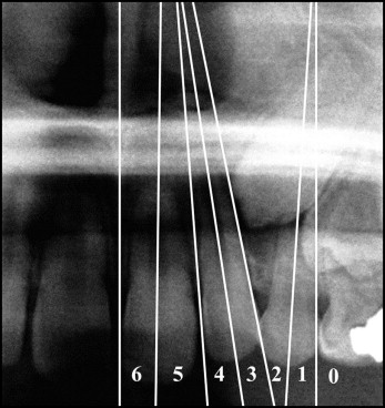
Fig 2 Sector designations used to assess maxillary canine and first premolar mesiodistal positions.
All measurements were performed twice with a 3-week interval by the same trained examiner (S.I.P.); all subsequent calculations were carried out using the arithmetic means between the 2, according to previous findings.
The following variables were also derived.
- 1.
The difference between the canine and premolar sectors on the first panoramic radiograph (S c-p ), used as an index of the intraosseous distance between the 2 teeth.
- 2.
The changes in canine and premolar inclinations between the 2 radiographic examinations (respectively, Δα and Δπ), obtained by subtracting their values on the first panoramic radiograph from the values on the second panoramic radiograph.
Statistical analysis
Repeated measurements allowed evaluation of intraobserver agreement, assessed using intraclass correlation coefficients (ICCs).
Since measurements from the right and left sides of the same subject might be correlated, to avoid the violation of independent assumptions for simple descriptive statistics and bivariate comparisons (such as t tests and correlations), data from both the left and right sides were used but analyzed separately. Differences in inclination values and sectors between the first and second panoramic radiographs were tested using, respectively, paired-samples t tests and Wilcoxon signed rank tests. Pearson correlation coefficients were calculated to determine whether there were any correlations between Δα and Δπ, Δα and S c-p , Δπ and S c-p , Δα and months between radiographs (ΔM), and Δπ and ΔM. A Pearson partial correlation analysis was performed to explore the relationship between Δα and Δπ after adjustment for S c-p and ΔM.
The combined data from the left and right sides were analyzed using a mixed linear regression model, with Δα as the dependent variable; Δπ, S c-p , ΔM, and side (right or left) as fixed effects; and patient-specific random effects for the intercept and the slope with respect to Δπ, accounting for individual differences.
All statistical analyses were performed with the statistical computing software R (Institute for Statistics and Mathematics, Wirtschaftsuniversität Wien, Vienna, Austria; available at http://www.R-project.org ). P <0.05 was set as the level for statistical significance.
Results
One hundred twenty-six panoramic radiographs from 63 subjects (32 boys, 31 girls; mean age at the first panoramic radiograph, 9.05 ± 0.79 years) were obtained. Two canine-premolar pairs were excluded from the final sample because of an excessive buccal displacement of the canine that did not allow a proper recognition of the long axis. Because the correlation between Δα and Δπ was the primary endpoint, the achieved sample size allowed detection of small to medium correlation effects of r = 0.25, assuming a 2-tailed alpha of 0.05 and a power level of 0.80.
The mean time interval between the first and the second panoramic radiographs was 18.7 ± 3.5 months.
The intraobserver agreement was excellent for both angular measurements (angle α: ICC, 0.997; angle π: ICC, 0.988) and sector location (S c : ICC, 1.000; S p : ICC, 0.983).
Significant differences between time points were detected for both canine and premolar inclinations to the midline. The premolar and left canine sectors varied statistically significantly too, whereas the right canine sector did not ( Table I ).
| n | Inclination (°) | Sector | |||||||
|---|---|---|---|---|---|---|---|---|---|
| T0 | T1 | t | P | T0 | T1 | Z | P | ||
| Maxillary canine | |||||||||
| R | 62 | 19.60 ± 10.07 | 14.67 ± 11.45 | 2.912 | <0.05 | 2.29 ± 0.71 | 2.08 ± 0.97 | −2.378 | <0.05 |
| L | 62 | 19.69 ± 10.98 | 14.66 ± 11.46 | 2.728 | <0.05 | 2.24 ± 0.64 | 2.11 ± 0.90 | −1.182 | NS |
| Maxillary first premolar | |||||||||
| R | 62 | 13.30 ± 7.69 | 8.94 ± 9.38 | 3.037 | <0.05 | 1.49 ± 0.59 | 0.56 ± 0.69 | −5.929 | <0.05 |
| L | 62 | 12.13 ± 7.89 | 8.68 ± 9.42 | 2.578 | <0.05 | 1.41 ± 0.61 | 0.65 ± 0.65 | −5.709 | <0.05 |
As reported in Table II , the Pearson correlation analysis showed a strong, positive correlation between Δα and Δπ, which proved to be statistically significant. Neither Δα nor Δπ showed significant correlations with S c-p and ΔM. After adjustment for S c-p and ΔM, the Pearson partial correlation coefficient between Δα and Δπ was larger than the zero-order correlation coefficient; this was because S c-p acted as a suppressor (ie, if it was not controlled, it suppressed the relationship between Δα and Δπ).
| Pearson correlation coefficient r ( P value) | Δα vs Δπ Pearson partial correlation coefficient r ( P value) | |||||||||||
|---|---|---|---|---|---|---|---|---|---|---|---|---|
| Δπ | S c-p | ΔM | Adj S c-p | Adj ΔM | Adj S c-p and ΔM | |||||||
| Right | Left | Right | Left | Right | Left | Right | Left | Right | Left | Right | Left | |
| Δα | 0.752 (0.000) |
0.772 (0.000) |
−0.220 NS |
−0.148 NS |
0.244 NS |
0.179 NS |
0.773 (0.000) |
0.793 (0.000) |
0.743 (0.000) |
0.765 (0.000) |
0.766 (0.000) |
0.785 (0.000) |
| Δπ | 0.004 NS |
0.068 NS |
0.156 NS |
0.191 NS |
||||||||
∗ Simple refers to Pearson correlation; partial refers to correlation after adjustment for S c-p , ΔM, or both.
Stay updated, free dental videos. Join our Telegram channel
VIDEdental - Online dental courses


